Key Takeaways
An acellular dermal matrix is a biologic or engineered scaffold used to support tissue repair and reconstruction. Its value depends on the material, the procedure, and the patient’s healing conditions.
- Decellularization removes most cells while retaining important extracellular matrix structure.
- The scaffold can support cellular ingrowth, vascularization, and gradual tissue remodeling.
- Applications include wound care, breast reconstruction, hernia repair, and soft-tissue reconstruction.
- Material source, thickness, strength, preservation, and regulatory status affect clinical selection.
- Benefits must be weighed against infection, seroma, inflammation, cost, and procedure-specific uncertainty.
What an acellular dermal matrix is
An acellular dermal matrix is a three-dimensional material used to support the repair or reconstruction of soft tissue. It may be derived from human or animal tissue, or produced from engineered materials. For you as a clinician or patient, the central idea is straightforward: the matrix provides structure while the body gradually responds to and remodels that structure. Its performance is not automatic; surgical technique, wound conditions, and host health all matter.
Definition and basic structure
A dermal matrix usually contains an extracellular framework made from proteins such as collagen, with other matrix components varying by source and processing method. The material is prepared so that many cellular elements are removed, while the remaining framework can be placed where additional tissue support is needed. It is not a replacement for living skin, and it does not function like a conventional skin graft.
The matrix may arrive as a sheet, mesh-like construct, or other shaped implant. Its intended role depends on the product and indication, so you should rely on the device labeling and the treating team’s plan rather than assuming that every matrix behaves in the same way.
How decellularization preserves the extracellular matrix
Decellularization uses physical, chemical, enzymatic, or combined processing methods to reduce cellular material and genetic remnants. The aim is to retain useful architecture and matrix molecules while limiting the immune response associated with living donor cells. The balance is delicate: aggressive processing may affect structure or biologic signals, while incomplete processing may leave more residual material than intended.
Quality assessment can therefore include testing for residual DNA, sterility, structural integrity, and other product-specific characteristics. These tests do not eliminate clinical uncertainty, but they help define what a particular matrix is designed and validated to do.
Common biologic and synthetic sources
Human dermis and animal tissues are common biologic sources. Animal-derived matrices may come from tissues such as porcine or bovine dermis, while synthetic options use polymers or other engineered materials. Each source brings different considerations involving mechanical behavior, processing, availability, host response, and regulatory classification.
A matrix based on natural tissue may retain aspects of native architecture, but that does not mean it is identical to the patient’s own dermis. Conversely, a synthetic material may offer more predictable physical properties without reproducing every biologic feature of native extracellular matrix.
How it differs from skin grafts and other surgical meshes
A skin graft transfers living or viable skin tissue to cover a wound, whereas an acellular dermal matrix primarily provides a scaffold for repair. A surgical mesh is generally selected for reinforcement and load-bearing, particularly in abdominal wall procedures, although some matrices may also contribute to soft-tissue support. The categories can overlap in practical use, but their composition and intended biologic roles differ.
For a broader clinical review of human acellular dermal matrix use, you can consult this reconstructive surgery review. It is still essential to distinguish general literature from the instructions and evidence for the specific material being considered.
How acellular dermal matrices work in the body
Once placed, a matrix interacts with the wound environment rather than simply sitting inertly in the body. Fluid movement, inflammatory signaling, cell migration, vascular growth, and mechanical forces all influence what happens next. The matrix may gradually become incorporated into surrounding tissue, although the rate and quality of incorporation vary considerably. Your surgeon will judge that process in the context of the operation and the patient’s healing course.
Scaffold function and tissue integration
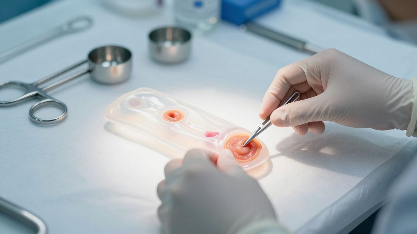
The matrix gives cells a temporary or lasting physical framework in a region where tissue has been lost, weakened, or surgically rearranged. It can help maintain a contour, cover exposed structures, reinforce a repair, or provide a surface over which new tissue develops. The scaffold is not the healing process itself; it is one component of a carefully managed repair environment.
Integration depends on close contact with a suitable wound bed. Excess fluid, dead space, contamination, or inadequate fixation can interfere with that contact and create conditions in which the matrix does not perform as intended.
Revascularization and cellular repopulation
After implantation, blood vessels and host cells may grow into the matrix when local conditions support that response. Revascularization can bring oxygen and nutrients, while repopulating cells contribute to the formation of new connective tissue. The pattern is influenced by the matrix’s porosity, thickness, processing, and placement, as well as the vascularity of the recipient site.
This is one reason surgeons pay attention to tissue quality and wound-bed preparation. A matrix placed into poorly perfused or infected tissue may face a very different biologic environment from one placed into clean, healthy tissue.
Remodeling over time
Remodeling means that the implanted material and the surrounding tissue change over time. Enzymes, fibroblasts, immune cells, and mechanical forces can alter the scaffold’s organization and strength. Some materials are intended to be gradually absorbed, while others are designed to remain for longer-term support.
You may notice that early postoperative findings do not predict the final result. Swelling, fluid collections, and immature scar tissue can change as healing proceeds, which is why clinical review and, when indicated, imaging are interpreted in relation to the timing of surgery.
Factors that influence incorporation
Incorporation is shaped by both the material and the host. Important variables include wound cleanliness, blood supply, tension, fixation, fluid control, smoking, diabetes, nutritional status, immune function, and the size of the reconstructed area.
The treating team may therefore focus on several practical priorities:
- preparing a viable, adequately vascularized wound bed;
- reducing contamination and managing dead space;
- selecting a size and thickness that fit the defect;
- securing the material without excessive tension;
- monitoring the wound for fluid, infection, or separation.
These measures do not guarantee incorporation, but they address conditions that commonly affect tissue repair. They also show why the same matrix can have different outcomes in different operations.
Major clinical applications
Acellular dermal matrices are used across reconstructive and wound-care settings, but their purpose changes with the anatomy and surgical goal. In one case, the matrix may reinforce an implant pocket; in another, it may provide coverage over a defect or support a weakened abdominal wall. Evidence is often procedure-specific, so results from one indication should not be transferred casually to another. Your surgeon should explain the expected role of the selected material in your operation.
Breast reconstruction and implant support
In implant-based breast reconstruction, an acellular matrix may help support the implant, define the pocket, and influence soft-tissue contour. It can be used as part of a staged or immediate reconstruction plan, depending on the patient’s anatomy and the surgeon’s technique. The decision also takes into account mastectomy skin quality, radiation history, infection risk, and the amount of available soft-tissue coverage.
Postoperative imaging can show expected matrix-related findings as well as complications. A radiologist who knows the surgical history can better distinguish normal postoperative appearances from seroma, hematoma, infection, fat necrosis, or recurrent disease.
Hernia and abdominal wall repair
For abdominal wall repair, a matrix may be considered when reinforcement or biologic tissue support is needed. The relevant questions include defect size, contamination, tension, tissue quality, recurrence risk, and the required mechanical strength. A material suitable for a small soft-tissue defect may not be appropriate for a large load-bearing abdominal wall reconstruction.
Because abdominal wall repairs experience repeated movement and pressure, fixation and positioning are especially important. The matrix is part of the repair strategy, not a substitute for sound surgical closure or management of factors that increase recurrence risk.
Wound coverage and soft-tissue reconstruction
Matrices may be used to support coverage of selected burns, chronic wounds, surgical defects, and areas of soft-tissue loss. In wound care, the wound bed must be assessed carefully for necrotic tissue, infection, perfusion, exudate, and the need for debridement. The matrix may then provide a structured surface that supports the body’s repair response.
For clinicians evaluating tissue-engineered wound products, CholeDerm provides product-specific guidance on application, wound-healing stages, and monitoring. That information should not be generalized to every acellular matrix, because indications, preparation, and handling vary by product.
Head and neck, oral, and maxillofacial procedures
In head and neck, oral, and maxillofacial surgery, matrices may support soft-tissue coverage, mucosal repair, or reconstruction around anatomically complex structures. The choice must account for saliva, bacterial exposure, movement, thin tissue planes, and the need to preserve speech, swallowing, facial movement, or appearance.
These procedures often require precise shaping and fixation. A material that is easy to handle in a broad wound may behave differently in a narrow oral or facial site, so the operative plan should be tailored to the defect rather than based only on the matrix’s general category.
Types and material characteristics
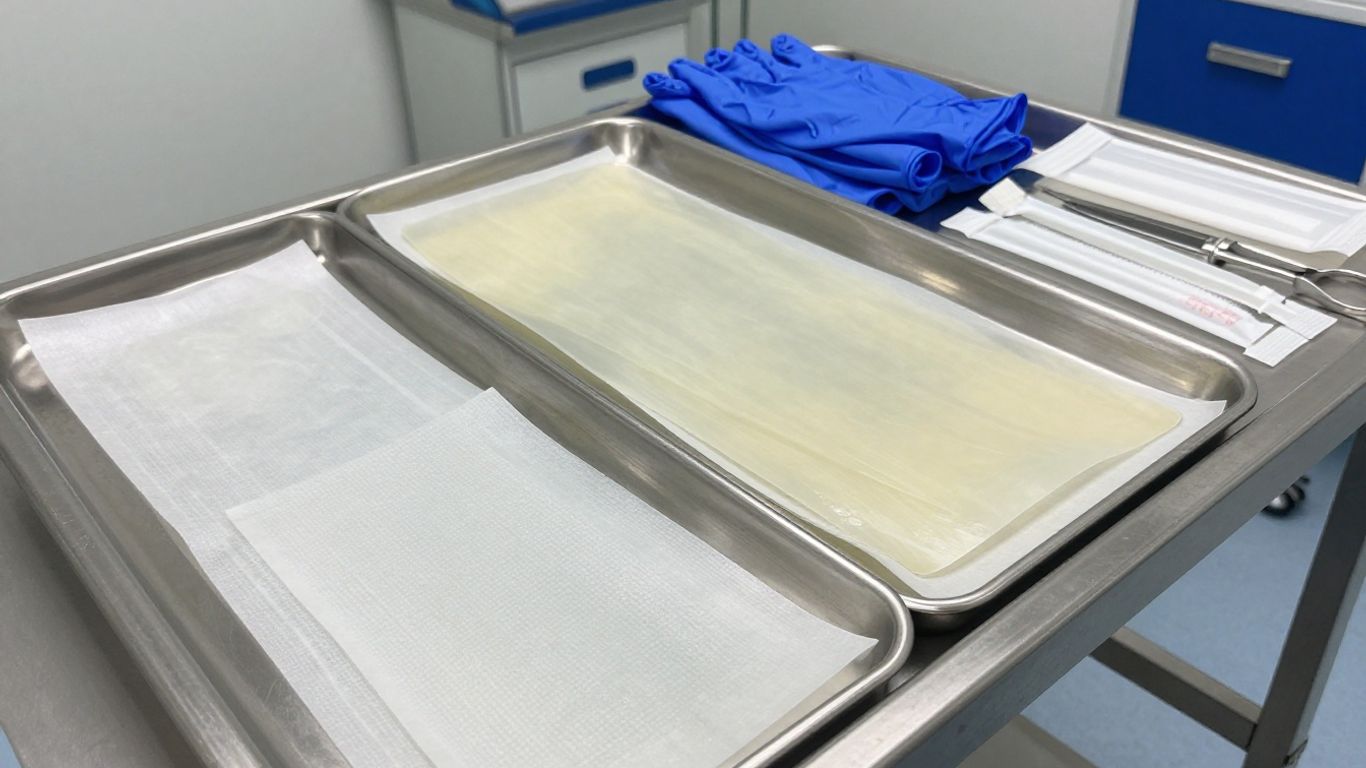
Not all acellular dermal matrices are interchangeable. Their source, processing, storage method, thickness, porosity, and mechanical properties can affect handling and clinical behavior. A product may be optimized for wound coverage, reinforcement, or tissue support, but its labeling defines the boundaries of appropriate use. Comparing products therefore requires more than comparing names or surface appearance.
Human, animal-derived, and synthetic options
Human-derived matrices are processed from donated human tissue, while animal-derived materials use tissues from sources such as porcine or bovine tissue. Synthetic matrices are manufactured from polymers or other engineered substances. The source may influence biologic composition, strength, degradation, immune considerations, supply, and cost.
Animal-tissue-derived biomaterials are also being developed for wound healing. Alicorn Medical, for example, describes CholeDerm as a tissue-engineered wound care product rather than presenting it as a universal replacement for every reconstructive matrix. Product-specific evidence and labeling remain the appropriate basis for clinical decisions.
Sterilization and preservation methods
Sterilization and preservation are central to safe storage and use. Products may be supplied dry, hydrated, frozen, or in another preserved state, and preparation before implantation can differ. Sterilization methods may affect collagen structure, residual chemicals, moisture, and mechanical behavior.
You should follow the manufacturer’s instructions for storage, rehydration, preparation, and handling. A matrix that has been stored incorrectly or prepared outside its validated process may not perform as expected, even if its visible appearance seems unchanged.
Thickness, porosity, and mechanical strength
Thickness can influence handling, coverage, fluid movement, and the time needed for tissue penetration. Porosity affects how cells and fluids move through the scaffold, while mechanical strength matters when the material must resist tension or support a repair. These properties must be considered together rather than treated as isolated advantages.
The following comparison illustrates the kinds of questions clinicians may ask during selection:
| Characteristic | Why it matters clinically | Questions for the surgical team |
|---|---|---|
| Thickness | Affects coverage, handling, and tissue penetration | Is the material suitable for this defect depth? |
| Porosity | Influences fluid movement and cellular access | Will the structure support the intended integration? |
| Mechanical strength | Determines resistance to tension and load | Does it match the forces at the repair site? |
| Preservation state | Affects storage and preparation | What preparation steps are validated? |
These characteristics do not independently predict a patient’s outcome. They become useful when matched to the anatomy, wound environment, fixation plan, and intended duration of support.
Permanent versus absorbable materials
Some matrices are intended to remain as longer-term structural support, while others are gradually degraded and replaced or remodeled by host tissue. Absorbable materials may be attractive when permanent foreign material is undesirable, but their strength and duration must suit the repair. Permanent materials may provide durable support but can carry different long-term considerations.
The distinction is not simply a matter of “natural” versus “synthetic.” A product’s actual degradation profile, clinical indication, and evidence should guide the discussion.
How clinicians select an acellular dermal matrix
Selection begins with the clinical problem, not with the material alone. Your care team considers the defect, the tissue bed, the expected forces, the desired duration of support, and the patient’s capacity to heal. Cost, availability, preparation time, and local regulatory requirements may also influence the final choice. A well-matched matrix still requires appropriate surgery and follow-up.
Matching the material to the procedure
The intended use should align with the matrix’s validated indication and physical characteristics. A wound-covering scaffold, an implant-support material, and an abdominal wall reinforcement product may have very different requirements. Clinicians also consider whether the material needs to be shaped, sutured, trimmed, hydrated, or combined with another reconstructive technique.
The most useful comparison is therefore procedure-specific. Asking whether a matrix is “good” in general is less informative than asking whether it is appropriate for the anatomy, tissue quality, and mechanical demands of a particular repair.
Patient health and wound-related considerations
Medical history can change the risk-benefit calculation. Diabetes, smoking, malnutrition, vascular disease, immunosuppression, prior radiation, infection, and previous surgery may affect vascularity and wound closure. The team may address modifiable risks before surgery or choose another strategy when the wound environment is unfavorable.
The patient’s goals matter as well. Coverage, durability, appearance, function, recovery time, and the possibility of additional procedures may carry different importance for different people.
Handling, sizing, and fixation requirements
A matrix must fit the defect and remain stable during early healing. Surgeons may trim it, overlap edges, place it in a particular plane, or secure it with sutures or other fixation methods. The instructions for use should define preparation and handling limits, including whether the material can be reshaped or stored after opening.
Small technical details can have practical consequences. Excessive tension, folding, poor contact, or unaddressed dead space may undermine an otherwise reasonable selection.
Regulatory status and manufacturer documentation
Regulatory authorization, labeling, instructions for use, contraindications, warnings, and post-market information should be reviewed before clinical use. Documentation also helps clarify the intended patient population, storage conditions, sterilization status, and evidence supporting the product. Alicorn Medical, for instance, describes its PrisTINE process and tissue-engineered product development in the context of biomaterials for wound healing; those descriptions do not replace product-specific regulatory documentation.
For clinicians, a careful records review is part of patient safety. For patients, it is reasonable to ask which product is being proposed, why it fits the procedure, and what alternatives have been considered.
What to expect during treatment
Treatment varies widely because an acellular dermal matrix can be used in different operations. You may encounter it during a planned reconstruction, wound-bed procedure, or revision surgery. The team will explain the operation, anesthesia, preparation, expected recovery, and potential complications. The matrix itself is only one part of that plan.
Preoperative assessment and surgical planning
Before surgery, clinicians assess the defect, surrounding tissue, blood supply, infection status, and expected mechanical demands. They may review imaging, photographs, prior operative reports, medication use, and conditions that affect healing. The plan often includes an alternative approach in case the wound or anatomy differs from expectations during surgery.
You should receive instructions about bathing, medications, fasting, smoking, nutrition, and postoperative support when relevant. Questions about the material, its source, and its expected role are appropriate before consent.
Placement and attachment techniques
During the procedure, the matrix is prepared according to its instructions and positioned against or within the target tissue. The surgeon may trim it to match the defect and secure it with sutures or another approved fixation method. Fluid control, tissue contact, and coverage of the intended area are important parts of placement.
The appearance of the matrix in the operating room does not by itself predict incorporation. Its outcome depends on the quality of the repair, the wound environment, and the patient’s subsequent healing.
Healing, monitoring, and follow-up imaging
Follow-up may include wound checks, dressing changes, assessment of pain and drainage, and evaluation of function or contour. Imaging is ordered when it can answer a specific clinical question, such as evaluating a collection or distinguishing expected postoperative findings from a complication. In reconstructed breasts, imaging interpretation can be improved when the radiologist knows the type and location of the implanted matrix.
Recovery timelines vary by procedure and by the amount of tissue repaired. Your instructions about activity, compression, dressings, and return visits should take priority over generalized timelines found online.
Signs of normal integration versus complications
Mild swelling, tenderness, bruising, and changes in firmness can occur during normal recovery, but the expected pattern depends on the operation. Increasing pain, spreading redness, fever, foul drainage, wound separation, sudden swelling, or persistent fluid should be reported promptly. These signs do not prove that the matrix has failed, but they warrant clinical assessment.
Do not remove dressings, drain fluid, or apply unapproved products without guidance. Early evaluation can help the team distinguish a manageable postoperative issue from infection, hematoma, seroma, or impaired wound healing.
Benefits, risks, and current evidence
Acellular dermal matrices can offer useful structural and biologic support, but they are not risk-free and do not eliminate the complexity of reconstruction. Evidence differs by product, indication, patient population, and study design. Some studies show potential benefits in selected settings, while others find trade-offs involving complications, cost, or uncertain long-term effects. A balanced discussion is more helpful than treating the material as universally superior.
Potential advantages in reconstructive surgery
Potential advantages include providing coverage, supporting tissue planes, helping maintain contour, and offering a framework for host tissue incorporation. In some situations, a matrix can expand the options available to the surgical team, particularly when local tissue is limited or additional support is needed.
The practical benefit is always contextual. A scaffold may be valuable when it solves a specific reconstructive problem, but its use should be connected to a clear surgical objective and a realistic recovery plan.
Infection, seroma, inflammation, and rejection-related concerns
Possible complications include infection, seroma, hematoma, inflammation, wound separation, chronic discomfort, poor incorporation, and recurrence of the original defect. Biologic matrices are processed to reduce cellular content, but processing does not make complications impossible. Immune and inflammatory responses can still occur, and contamination can compromise the repair.
Patients with impaired healing may face higher risks, although no single risk factor determines the outcome. Prompt reporting of concerning symptoms gives the clinical team a better opportunity to investigate and treat problems.
Limitations and procedure-specific trade-offs
Limitations may include cost, variable availability, preparation requirements, uncertain comparative benefit, and the need for additional surgery if the reconstruction fails. A matrix may also provide less mechanical support than a repair requires, or remain stronger or longer-lasting than is desirable for a particular tissue plane.
The relevant comparison is not always between two matrices. Depending on the case, alternatives may include primary closure, autologous tissue, a skin graft, a synthetic mesh, negative-pressure therapy, or staged reconstruction. Your surgeon should explain why one approach is preferred for your situation.
Interpreting clinical studies and long-term outcomes
When reading research, look for the indication, comparator, follow-up period, sample size, complication definitions, and whether the product studied matches the one being considered. A favorable result in wound coverage may not establish benefit in breast reconstruction or hernia repair. Manufacturer-supported evidence can be useful, but independent studies and transparent reporting also matter.
Long-term outcomes should include more than early closure. Recurrence, infection, reoperation, function, appearance, patient-reported outcomes, and durability may all be relevant. The strongest decision combines published evidence with clinical judgment and the particulars of your case.
Conclusion
An acellular dermal matrix is best understood as a procedure-specific scaffold that can support tissue repair, reconstruction, or wound coverage. Its source, structure, processing, and intended duration of support must match the patient and the surgical goal. By discussing the evidence, risks, alternatives, and follow-up plan with your care team, you can approach the decision with a clearer and more realistic understanding of what the material can and cannot do.
Frequently Asked Questions
What is an acellular dermal matrix?
It is a scaffold made from processed biologic tissue or engineered material, designed to support soft-tissue repair while reducing or removing cellular components.
Is an acellular dermal matrix the same as a skin graft?
No. A skin graft transfers skin tissue, while an acellular dermal matrix mainly provides a structural framework that the body may incorporate and remodel.
Where are acellular dermal matrices used?
They may be used in wound coverage, breast reconstruction, abdominal wall repair, and selected head and neck, oral, or maxillofacial procedures.
Does the body absorb an acellular dermal matrix?
That depends on the material. Some matrices are gradually degraded and remodeled, while others are intended to provide longer-term structural support.
What affects whether a matrix incorporates successfully?
Blood supply, wound cleanliness, fixation, fluid control, tissue quality, smoking, diabetes, nutrition, immune function, and the size and location of the repair can all influence incorporation.
What complications can occur?
Potential complications include infection, seroma, hematoma, inflammation, wound separation, poor incorporation, persistent pain, and recurrence of the original defect.
How should you choose an acellular dermal matrix?
The choice should be based on the procedure, wound environment, required mechanical support, patient health, product labeling, regulatory documentation, and the treating clinician’s experience.