
Key Takeaways
Elastin is one part of a larger biological system that helps skin move, resist stress, and recover after injury.
-
Elastin gives skin recoil and flexibility, while collagen provides much of its tensile support.
-
Elastic fibers participate in the extracellular matrix, where cells receive structural and biochemical cues.
-
Injury and inflammation can degrade elastin and leave scar tissue with poorly organized elastic fibers.
-
Age, ultraviolet exposure, diabetes, infection, and poor circulation can complicate elastic-fiber remodeling.
-
Regenerative scaffolds may help organize repair, but preclinical findings should not be treated as proof of human benefit.
What elastin is and how it supports healthy skin
The Role of Elastin in Skin Repair begins with a simple distinction: elastin is not the same protein as collagen. It is a stretchy structural protein embedded within elastic fibers, allowing skin to extend and then recoil. Its behavior depends on how it is assembled and how it interacts with the surrounding extracellular matrix.
If you picture skin as a living composite, collagen supplies much of the load-bearing framework while elastin helps that framework accommodate movement. This balance matters whenever you bend a joint, make a facial expression, or place tension across a healing wound. You can also read a broader overview of elastin structure and function for context beyond the skin.
The structure and organization of elastic fibers
Elastic fibers generally contain a central elastin-rich component surrounded by microfibrillar proteins. Tropoelastin molecules are assembled and cross-linked into a durable network, while the associated microfibrils help guide organization and connect the fibers with neighboring matrix components. The result is not a loose collection of protein strands but an integrated, three-dimensional system.
That organization gives elastic fibers their resilience. When mechanical force stretches the skin, the fibers deform; when the force is released, their molecular structure helps drive recoil. With injury, the body must rebuild more than elastin itself—it must restore orientation, attachment, and the relationship between fibers and other matrix materials.
Where elastin is found in the skin
Elastin is present mainly in the dermis, where it is distributed through networks that support skin movement and recoil. It is found around cells and alongside collagen fibers, blood vessels, and other extracellular components. Its abundance and arrangement vary by location, age, and exposure to mechanical or environmental stress.
The superficial and deeper dermal layers do not experience identical forces, so their elastic networks are not identical either. This helps explain why an injury can close at the surface while deeper tissue remains stiff or mechanically fragile. A closed wound is therefore not necessarily a fully remodeled wound.
How elastin differs from collagen
Collagen and elastin are both structural proteins, but they solve different mechanical problems. Collagen resists pulling forces and helps maintain tissue strength; elastin tolerates repeated stretch and supports recoil. Their functions overlap within the matrix, yet replacing one with the other would not recreate healthy skin mechanics.
This distinction is useful when you evaluate biomaterials or wound treatments. A material described only as collagen-rich may provide a structural framework without reproducing the full range of elastic, adhesive, and signaling functions found in native tissue. The practical question is not whether one protein is superior, but whether the matrix preserves a suitable combination of components.
Why elasticity matters for skin strength and flexibility
Elasticity allows skin to move without tearing every time its shape changes. It also helps distribute local forces across a broader area, which can be relevant around joints, incisions, and wounds exposed to repeated motion. Mechanical continuity matters because a fragile or rigid repair may tolerate stillness but struggle under normal activity.
You can think of elasticity as one contributor to functional healing rather than a cosmetic property alone. When the repaired tissue gradually regains flexibility, movement may become more comfortable and the risk of recurrent stress at the wound margin may be reduced, although clinical outcomes depend on many additional factors.
Elastin’s role throughout the wound-healing process
Wound healing proceeds through overlapping phases rather than a neat sequence of isolated steps. Hemostasis and inflammation stabilize the injury, proliferation rebuilds coverage and matrix, and remodeling gradually changes the strength and flexibility of the repair. Elastin is involved most clearly in the later mechanical organization of tissue, but events in the early phases can determine whether elastic fibers are ultimately preserved or disordered.
During repair, fibroblasts, keratinocytes, endothelial cells, immune cells, and matrix proteins communicate continuously. Their activity is influenced by oxygen, moisture, cytokines, enzymes, mechanical loading, and the condition of the wound bed. That is why elastin cannot be considered separately from the broader healing environment.
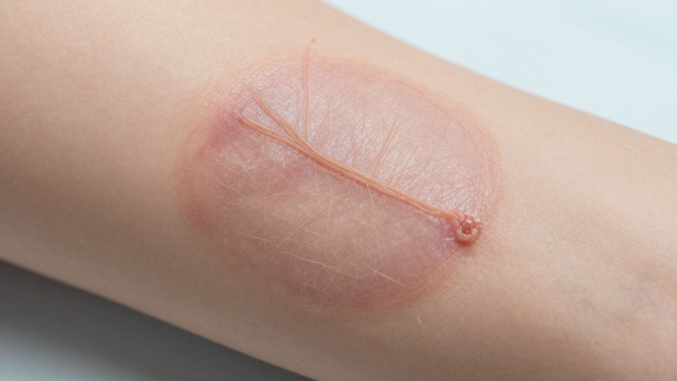
Elastic fibers during inflammation and wound stabilization
Early inflammation helps remove damaged material and defend against microbes, but inflammatory enzymes can also break down matrix proteins. Elastin is relatively durable, yet it is not protected from prolonged protease activity or oxidative stress. The initial clot and provisional matrix stabilize the wound, while immune cells begin shaping the conditions for later tissue formation.
A short, controlled inflammatory response may support repair. Persistent inflammation, by contrast, can prolong matrix degradation and prevent the transition toward organized remodeling. Clinically, this makes infection control and attention to the wound environment relevant not only to closure but also to the quality of the eventual tissue.
Fibroblast activity and new extracellular matrix formation
Fibroblasts enter or become active within the wound and produce provisional matrix components, including collagen and adhesive proteins. They also respond to growth factors and mechanical signals that influence migration, proliferation, and matrix organization. New elastin production is generally slower and more limited than early collagen deposition, so elastic restoration often lags behind surface closure.
As fibroblasts mature and the matrix changes, the balance between synthesis and degradation becomes important. A scaffold or wound interface that supports cell attachment may help cells occupy the repair site, but it cannot guarantee normal elastic-fiber formation. The behavior of the host cells and the surrounding biology remain decisive.
Re-epithelialization and restoration of skin movement
Keratinocytes migrate across the wound to restore the epidermal barrier. This protects the underlying repair from fluid loss and external exposure, but it does not by itself recreate the dermal elastic network. You may therefore see rapid epithelial coverage while deeper layers are still immature.
Movement places changing forces on newly covered skin. Gradual return to ordinary activity, guided by clinical judgment, allows the repair to adapt without excessive strain. The restored surface and the restored mechanical function are related milestones, not interchangeable ones.
Remodeling and the return of tissue flexibility
Remodeling can continue for months as collagen is reorganized, matrix components are degraded or replaced, and the tissue responds to mechanical loading. Elastic fibers may be newly deposited, but their arrangement can remain different from uninjured skin. Scar pliability reflects this complex history rather than elastin quantity alone.
A useful way to assess progress is to consider several outcomes together: closure, inflammation, tissue softness, range of motion, and symptoms. Improvement in one does not prove that every layer has returned to its original state. This broader view supports more realistic expectations for repair.
How elastin interacts with the extracellular matrix
The extracellular matrix is more than a passive scaffold. It provides physical support, binds water, presents attachment sites, and carries signals that influence how cells behave. Elastin sits within this network, so its function depends on the surrounding molecules and on the architecture that places them in relation to one another.
This is why a biologically complex matrix can behave differently from a purified single-protein material. Composition, fiber arrangement, porosity, degradation, and retained signaling molecules all affect how a wound responds. In tissue engineering, the challenge is to preserve useful biology while creating a material that can be handled safely and consistently.
The relationship between elastin, collagen, and glycosaminoglycans
Collagen provides much of the matrix framework, elastin contributes recoil, and glycosaminoglycans attract water and help organize the local biochemical environment. Proteoglycans associated with glycosaminoglycans can also influence growth-factor presentation and cell behavior. Together, these components form a hydrated, mechanically varied matrix rather than a single uniform material.
Their interaction matters during repair because hydration affects diffusion, cell movement, and tissue softness. A matrix that retains several native components may offer a closer approximation of the wound’s biological setting than one that contains only a concentrated structural protein. The result still depends on processing and host response.
Fibronectin and other proteins that support cell attachment
Fibronectin helps cells attach to and migrate across provisional or engineered matrix. Other proteins, including proteoglycans and basement-membrane-associated molecules, provide additional binding and organizational cues. These molecules can influence how fibroblasts and other repair cells spread, orient, and interact with the scaffold.
The distinction between a structural material and a biologically active matrix is useful here. A structure may occupy space, while a matrix with retained adhesion proteins can also provide recognizable interfaces for cells. Neither property alone guarantees regeneration, but both may be relevant to a healing wound.
Elastin-derived signals during tissue repair
Fragments released when elastin is degraded can act as biological signals, sometimes called elastin-derived peptides. Their effects depend on fragment size, concentration, receptors, cell type, and the inflammatory context. In a controlled repair process, matrix breakdown can help coordinate turnover; in chronic inflammation, the same process may contribute to persistent signaling and dysregulated remodeling.
For that reason, elastin degradation is not simply a matter of losing mechanical recoil. It can also alter the messages presented to immune and stromal cells. Researchers continue to study how these signals affect migration, inflammation, vascular responses, and matrix production.
Why preserving native matrix architecture may matter
The location of a molecule can be as important as its presence. Native fibers, pores, adhesive proteins, and growth-factor-binding sites create spatial relationships that are difficult to reproduce after aggressive processing. Preserving those relationships may help cells encounter a more familiar repair environment.
The principle is reflected in research on porcine cholecyst-derived matrices. CholeDerm® is described in the supplied preclinical material as retaining native collagen, elastin, glycosaminoglycans, and growth factors, with a thin, porous, mesh-like structure. These are material characteristics, not proof that every wound will regenerate normal elastic tissue.
What happens to elastin after skin injury
Injury changes the matrix immediately. Damaged elastic fibers may be fragmented, displaced, or exposed to inflammatory conditions that accelerate degradation. The body then builds a provisional matrix that is useful for closure but not necessarily equivalent to the original dermal architecture.
The final result depends on the wound’s depth, size, location, contamination, blood supply, mechanical stress, and duration of inflammation. A small acute wound may remodel relatively well, while a chronic or infected wound can remain trapped in a cycle of breakdown and incomplete repair.
Elastin degradation caused by inflammation and enzymes
Neutrophils, macrophages, and other cells release proteases and reactive molecules during inflammation. These help clear damaged tissue and microbes, but excessive or prolonged activity can damage elastin and neighboring matrix proteins. The balance between proteases and their inhibitors is therefore part of the wound’s biochemical stability.
When elastin is fragmented, the tissue may lose some recoil and may also generate breakdown signals. The body can replace matrix material, but adult skin does not always recreate the precise fiber network present before injury. This gap between replacement and restoration helps explain why scars often feel different.
Why scar tissue often contains disorganized elastic fibers
Scar formation prioritizes rapid closure and tensile reinforcement. Collagen is deposited and later reorganized, but elastic fibers may be sparse, irregularly oriented, or distributed differently from those in uninjured dermis. The scar can therefore be closed and strong while remaining tight, raised, depressed, or less mobile.
Scar quality changes over time, and gentle movement or clinical interventions may be appropriate in selected cases. Still, no single action can guarantee restoration of the original elastic architecture. Evaluation should consider the scar’s appearance, symptoms, function, and maturity.
Differences between acute wounds and chronic wounds
Acute wounds usually move through inflammation, proliferation, and remodeling with a recognizable change in cellular activity. Chronic wounds may show persistent inflammation, excess protease activity, impaired angiogenesis, microbial burden, or repeated mechanical disruption. These conditions make it harder for the matrix to progress from provisional repair to organized remodeling.
The contrast can be summarized by the dominant challenge in each setting:
|
Wound context |
Matrix challenge |
Likely effect on elastin-related repair |
|---|---|---|
|
Acute, clean wound |
Temporary inflammation and matrix disruption |
Remodeling may proceed if closure and perfusion are adequate |
|
Infected wound |
Persistent inflammatory and enzymatic activity |
Ongoing degradation can delay organization |
|
Diabetic wound |
Impaired vascular and cellular responses |
Slower closure and incomplete matrix maturation |
|
Ischemic wound |
Limited oxygen and nutrient delivery |
Fibroblast activity and remodeling may be constrained |
This comparison is a guide rather than a prediction for an individual patient. It highlights why treating the underlying wound condition is as important as covering the surface.
How diabetes, infection, and poor circulation can affect remodeling
Diabetes can affect immune function, angiogenesis, sensation, and cellular metabolism, while infection can sustain inflammatory signaling and increase tissue damage. Poor arterial or venous circulation can limit oxygen delivery or promote edema, both of which may hinder fibroblast activity and matrix turnover. Several factors often occur together rather than in isolation.
A thorough assessment should therefore consider diabetes control, vascular status, pressure, neuropathy, infection, nutrition, and medication history. Addressing these drivers can create better conditions for remodeling, although it cannot guarantee that the original elastic-fiber pattern will return.
Elastin, aging, and impaired skin repair
Aging changes the amount, organization, and turnover of dermal matrix proteins. The skin may become thinner, less resilient, and slower to respond to injury, while decades of environmental exposure add further stress. These processes are gradual, but they become visible when tissue is asked to heal or tolerate repeated movement.
Your repair capacity is influenced by both intrinsic aging and external damage. The distinction matters because ultraviolet exposure, smoking, inflammation, and illness may accelerate matrix injury beyond chronological age alone. A practical overview of elastin and visible skin aging can help connect these mechanisms with everyday skin changes.
How elastin production changes with age
Elastin synthesis and turnover change with age, and mature elastic fibers are not readily replaced once they are damaged. Older dermis may contain fragmented or poorly organized fibers alongside altered collagen. Fibroblasts can also become less responsive to signals that previously supported matrix production.
These changes do not mean that older skin cannot heal. They mean that the repair may be slower, less elastic, or more prone to persistent remodeling. Clinical planning should account for tissue quality rather than relying on age as a stand-alone explanation.
Photoaging and ultraviolet-related elastic fiber damage
Ultraviolet radiation can promote oxidative stress, inflammatory signaling, and enzyme activity that damages dermal matrix. Chronic exposure is associated with abnormal accumulations of elastic material and disrupted fiber organization, a pattern often called solar elastosis. The affected skin may look lax while also feeling less supple.
Protecting healing and intact skin from ultraviolet exposure is therefore relevant to long-term matrix preservation. Sun protection does not reverse established damage, but it can reduce one continuing source of stress during recovery.
The effect of oxidative stress and chronic inflammation
Reactive oxygen species can modify proteins and affect cellular signaling. Persistent low-grade inflammation may also keep matrix-degrading enzymes active and impair the coordinated transition toward repair. Together, these influences can change both the quality of newly produced matrix and the survival of existing fibers.
The skin’s antioxidant and inflammatory systems are connected to general health, medication use, infection, and environmental exposures. A wound that appears locally modest may still heal poorly when systemic or repeated inflammatory pressures are present.
Why aged skin may heal more slowly and lose flexibility
Older skin often has reduced thickness, altered blood supply, slower cell migration, and a less responsive extracellular matrix. These factors can delay epithelial coverage and prolong remodeling. When scar tissue forms, limited elastic-fiber replacement may leave the area tighter or less adaptable to motion.
You should interpret flexibility as a functional outcome, not merely a measure of appearance. Pain, tightness, range of motion, and the ability to tolerate ordinary activity can reveal problems that are not obvious from closure alone.
How regenerative materials may support elastin-related repair
Regenerative materials are designed to do more than cover a wound. They may provide a temporary structure, retain biochemical cues, manage the interface with the wound bed, and support host-cell migration. Whether they improve elastin-related repair depends on how well their composition and architecture fit the biological problem.
A scaffold does not replace the patient’s cells or eliminate the need for wound-bed preparation. It works within a broader system that includes perfusion, infection control, mechanical protection, and appropriate clinical monitoring. Evidence should therefore be read with attention to material, model, and outcome.
Natural extracellular matrix scaffolds as repair templates
A natural extracellular matrix scaffold can offer collagen, elastin, glycosaminoglycans, adhesive proteins, and other molecules in a three-dimensional arrangement. Cells may attach to the scaffold, migrate through it, and respond to retained signals as the material is remodeled. This makes the scaffold a potential repair template rather than a simple physical covering.
The template concept should not be confused with directing every cell toward a predetermined outcome. Host biology remains variable, and degradation must be balanced with the pace of new tissue formation. A scaffold that disappears too quickly may lose support, while one that persists too long may interfere with normal remodeling.
Elastin retention in decellularized biomaterials
Decellularization aims to reduce cellular remnants while preserving useful extracellular components. Processing choices matter: harsh detergents or enzymes may remove or alter proteins, whereas gentler approaches may better retain native architecture but require careful validation for safety and consistency.
Research behind CholeDerm® describes a non-detergent, non-enzymatic decellularization approach and reports preservation of collagen, elastin, glycosaminoglycans, and growth factors in the evaluated material. The same evidence base also distinguishes research findings from commercial claims, so retention of components should not be presented as a guaranteed clinical outcome.
Evidence from wound and tissue-engineering models
Preclinical studies can show how a material behaves before human use. In the supplied research, cholecyst-derived matrices were investigated in rabbit full-thickness wounds, burn models, diabetic-rat wounds, naturally occurring dog wounds, and soft-tissue models. Reported findings included granulation, epithelialization, angiogenesis, scaffold integration, and organized remodeling in the evaluated models.
For clinicians and researchers, the model is part of the result. A diabetic-rat study does not establish effectiveness in people with diabetes, and a formulation modified for research is not automatically the same as a commercial product. Careful reading preserves the useful signal without overstating translation.
Interpreting preclinical findings without overstating clinical benefits
Preclinical evidence is valuable for identifying mechanisms, safety questions, and design directions. It cannot by itself establish human efficacy, comparative superiority, or an appropriate indication. Those conclusions require suitable clinical evidence, product documentation, and professional judgment.
When you review a study, ask four questions: What material was tested? In which model? Which outcome was measured? How long was follow-up? This habit helps separate a promising biological observation from a clinical claim.
Supporting healthy elastin function during skin repair
You cannot control every aspect of elastic-fiber remodeling, but you can support the conditions in which repair occurs. Protecting the wound, maintaining a suitable moisture balance, treating infection, and managing systemic risks all influence the matrix environment. These measures complement—not replace—assessment by a qualified clinician.
The most useful approach is usually consistent and unglamorous. A wound should be reassessed when its appearance, drainage, pain, odor, or function changes, and treatment should be adjusted when progress stalls. Elastin is part of the biology, not a reason to overlook the basics of wound care.
Protecting healing skin from ultraviolet exposure
Newly healed skin is vulnerable to ultraviolet-related inflammation and pigment changes. Covering the area and using clinician-approved sun protection can reduce exposure while the barrier and deeper matrix continue to mature. Protection is especially relevant when a scar remains pink, thin, or easily irritated.
Sun avoidance should be practical rather than absolute. Clothing, shade, and appropriately used sunscreen can work together, with product choice guided by the wound’s stage and clinical advice.
Maintaining a balanced, moist wound environment
A wound that is excessively dry may impair cell migration, while excess fluid can macerate surrounding skin and increase local complications. Dressings are selected according to wound depth, exudate, tissue condition, contamination, and the need for inspection. The goal is a stable interface that protects new tissue without trapping harmful conditions.
Key aspects of a supportive environment include:
-
Adequate moisture for epithelial migration without surrounding maceration.
-
Control of exudate so fluid does not accumulate at the wound edge.
-
Low-adherence contact with fragile granulation or epithelial tissue.
-
Protection from friction, pressure, and repeated mechanical disruption.
These principles support the wound as a whole. They do not selectively increase elastin, but they can reduce avoidable obstacles to the cells and matrix involved in repair.
Managing inflammation, infection, and underlying conditions
Persistent inflammation should prompt a search for causes such as infection, necrotic tissue, pressure, foreign material, uncontrolled diabetes, or impaired perfusion. Managing these factors can help the wound leave the inflammatory phase and enter a more constructive repair state. The correct intervention depends on the wound and the patient rather than on elastin biology alone.
Nutrition, vascular assessment, off-loading, glucose management, and appropriate debridement may all be relevant in selected cases. A biologic scaffold or advanced dressing is most sensibly considered as part of this coordinated plan, not as a substitute for it.
When clinical evaluation is important for delayed or abnormal healing
Seek clinical evaluation when a wound is enlarging, increasingly painful, foul-smelling, draining heavily, bleeding unexpectedly, or failing to show gradual improvement. Fever, spreading redness, discoloration, new numbness, or loss of function also deserves prompt attention. People with diabetes, vascular disease, immune suppression, or neuropathy may need earlier review because complications can progress quietly.
A clinician can assess perfusion, infection, pressure, tissue viability, and the need for imaging or specialist care. Early evaluation may protect the remaining matrix and reduce the duration of damaging inflammation.
Conclusion
Elastin helps skin accommodate movement and recover its shape, but its role in repair is inseparable from collagen, glycosaminoglycans, adhesive proteins, immune signaling, and tissue architecture. Injury, aging, ultraviolet exposure, and chronic disease can disrupt this network, while regenerative materials may offer useful templates in carefully studied settings. The most reliable path to better healing remains a complete plan that addresses the wound environment, the patient’s underlying conditions, and the limits of current evidence.
Frequently Asked Questions
What is elastin’s main function in skin?
Elastin helps skin stretch and recoil, contributing to flexibility and mechanical resilience. It works alongside collagen and other extracellular matrix components rather than functioning alone.
Is elastin the same as collagen?
No. Collagen provides much of the skin’s tensile framework, while elastin supports stretch and recoil. Healthy dermal mechanics depend on the interaction of both proteins.
Does a closed wound have fully restored elastin?
Not necessarily. Surface closure can occur before deeper matrix remodeling is complete, and scar tissue may contain elastic fibers that are reduced or organized differently from those in uninjured skin.
Why can chronic wounds have poor elastin remodeling?
Chronic wounds may remain inflamed and can have infection, excess enzyme activity, poor circulation, diabetes-related dysfunction, or repeated mechanical stress. These conditions interfere with organized matrix turnover.
Can aging affect elastin-related repair?
Yes. Aging can change elastin production, fiber organization, fibroblast responsiveness, skin thickness, and blood supply. As a result, repair may be slower and the healed tissue may be less flexible.
Can ultraviolet exposure damage elastin?
Repeated ultraviolet exposure can promote oxidative stress, inflammation, and abnormal elastic-fiber remodeling. Protecting healing and intact skin from ultraviolet radiation helps limit ongoing damage.
When should you ask a clinician about a wound?
Ask for clinical evaluation if a wound worsens, becomes increasingly painful, develops spreading redness or odor, drains heavily, loses function, or fails to improve. Earlier assessment is especially important when diabetes, vascular disease, neuropathy, or immune problems are present.