
Key Takeaways
Glycosaminoglycans help turn the extracellular matrix from passive scaffolding into an active environment for repair.
- GAGs hold water and influence tissue hydration.
- Their charge helps organize growth-factor signaling.
- GAGs affect cell migration, proliferation and immune behavior.
- Preserving native GAGs can support the biological function of ECM scaffolds.
- Cholecyst-derived ECM offers a useful model for combining structure with regenerative signals.
What glycosaminoglycans are and where they fit in the extracellular matrix
If you want to understand why glycosaminoglycans matter in regenerative medicine, start with the extracellular matrix, or ECM. The ECM is not simply the material between cells; it is a changing, information-rich environment that influences how cells attach, move and behave. GAGs are one part of that environment, working alongside proteins, water and signaling molecules. A helpful overview of glycosaminoglycans places these molecules at the center of current tissue-engineering research.
The structure of glycosaminoglycans and proteoglycans
GAGs are long, unbranched carbohydrate chains built from repeating disaccharide units. Many are attached to a core protein, creating proteoglycans, while hyaluronic acid usually exists as a free polysaccharide rather than a classic proteoglycan side chain. The resulting molecules occupy space, bind water and interact with proteins in the surrounding matrix. Proteoglycans therefore act as more than structural fillers: they help organize a hydrated three-dimensional environment.
Major types, including hyaluronic acid, heparan sulfate and chondroitin sulfate
The major GAG families differ in their sugar composition, sulfation and biological interactions. Hyaluronic acid contributes to hydration and can support cell movement; heparan sulfate often participates in growth-factor and cell-surface signaling; and chondroitin sulfate contributes to matrix organization and tissue mechanics. Dermatan sulfate, keratan sulfate and heparin add further functional diversity. You can explore the broader GAG classification when comparing these families.
How GAGs differ from collagen and other ECM components
Collagen provides much of the tensile framework of connective tissue, while elastin supports recoil and adhesion proteins help cells attach to the matrix. GAGs work differently. They occupy hydrated spaces and regulate molecular traffic around the fibers and cells, so the ECM can provide both physical support and biochemical instruction. That distinction is why collagen-rich scaffolds may still behave differently depending on which non-collagenous components remain.
Why their negative charge makes them biologically active
Many GAGs carry substantial negative charge because of sulfate and carboxyl groups along their chains. This charge attracts water and positively charged regions of proteins, allowing GAGs to bind, concentrate or restrict signaling molecules. The effect depends on chain length, sulfation pattern, location and degradation. In practice, the same chemical feature that supports hydration can also shape how cells experience growth factors and inflammatory cues.
How GAGs regulate the regenerative microenvironment
A wound is a moving biochemical system rather than a static defect. As fluid balance, inflammation and cell populations change, the ECM helps coordinate what happens next. GAGs contribute by creating hydrated spaces, presenting signals and modifying how cells encounter one another. Their activity is contextual, which means preservation of the native matrix may matter as much as the presence of any single molecule.
Retaining water and maintaining tissue hydration
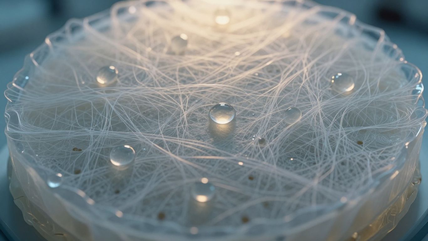
GAGs attract and retain water within the ECM, helping maintain the hydrated conditions that support diffusion and cell movement. This does not mean that more moisture is always better in a wound; excess fluid and poor exudate control can be harmful. Rather, a balanced hydrated matrix gives cells a workable environment for migration and matrix deposition. Hyaluronic acid and sulfated GAGs are especially relevant to this physical aspect of repair.
Binding growth factors and controlling their availability
GAGs can bind growth factors and other signaling proteins, influencing where those signals remain and how quickly they are released. This may protect some molecules from rapid diffusion while presenting them near responsive cells. The result is not a simple on-or-off switch. Binding strength, enzymatic remodeling and local tissue conditions all help determine whether a signal is retained, released or altered.
Supporting cell adhesion, migration and proliferation
Cells respond to both the chemistry and the geometry of their matrix. GAG-rich regions can affect integrin-mediated adhesion indirectly, support migration through hydrated spaces and change the presentation of factors that encourage proliferation. Researchers therefore study GAG-containing constructs alongside collagen, fibronectin and other adhesion molecules. A matrix that preserves several of these cues may provide a more realistic substrate than one reduced to a single purified component.
Influencing inflammation and macrophage behavior
GAG fragments and intact matrix molecules can influence inflammatory signaling, although the outcome depends on their size, concentration and surrounding molecules. Macrophages may shift between inflammatory and reparative states as the wound evolves, and the ECM participates in that conversation. This is one reason regenerative scaffolds are evaluated for host-material interaction, not only for mechanical strength. The biology should be interpreted as modulation rather than a promise of inflammation-free healing.
The role of GAGs in wound healing and tissue repair
Wound repair proceeds through overlapping inflammatory, proliferative and remodeling phases. GAGs participate across that sequence, but their contribution changes as the matrix is broken down and rebuilt. You can think of them as part of the tissue’s signaling infrastructure: they help regulate the conditions in which immune cells, endothelial cells, keratinocytes and fibroblasts operate. This complements broader work on tissue regeneration, where matrix-cell interactions are studied as a connected system.
Supporting the inflammatory and proliferative phases
Early inflammation is necessary for debris clearance and defense, but it must eventually give way to constructive repair. GAGs can influence the retention and movement of cytokines and growth factors during this transition. As the proliferative phase begins, their hydrated environment supports cell movement and provisional matrix formation. Their role is therefore less about accelerating one isolated step and more about helping the wound change state in an orderly way.
Promoting granulation tissue and angiogenesis
Granulation tissue requires fibroblasts, new vessels, provisional matrix and a controlled immune response. GAGs can influence angiogenic signaling by binding or presenting factors near endothelial cells. That mechanism is relevant to vascular biomaterial design, where vascular tissue regeneration depends on combining suitable architecture, degradation and biochemical cues. Still, angiogenesis in a material is a measured biological outcome, not an automatic consequence of adding a GAG.
Helping keratinocytes and fibroblasts coordinate repair
Keratinocytes need a surface that permits migration across the wound, while fibroblasts populate deeper regions and produce new matrix. Hydration, adhesion sites and growth-factor presentation affect both cell types, but not in identical ways. A biologically complex ECM can provide several overlapping cues, allowing epithelial and stromal cells to respond to a changing local environment. In a chronic wound, that coordination may be disrupted by persistent inflammation, impaired vascularization or excessive protease activity.
Contributing to collagen organization and tissue remodeling
During remodeling, early matrix is gradually replaced and reorganized. GAGs can affect collagen fibril assembly, matrix hydration and the activity of enzymes that reshape tissue. The desired result is not simply more collagen; it is a better-organized matrix with appropriate mechanical and biological behavior. This distinction matters when evaluating whether a scaffold supports constructive remodeling rather than only short-term wound closure.
Why GAG preservation matters in biological scaffolds
Processing a biological tissue into a scaffold can change its chemistry, structure and degradation behavior. Detergents, enzymes, washing steps, drying and sterilization-related conditions may remove or modify matrix components. A scaffold can retain a recognizable collagen structure while losing some of the molecules that made the original tissue biologically instructive. For this reason, scaffold characterization should consider architecture and biomolecule retention together.
How processing can alter native ECM biomolecules
Decellularization aims to reduce cellular material and immunogenic residues while preserving useful ECM components. Those goals can conflict: harsher processing may improve removal of cells but also disrupt proteins, GAGs or growth-factor activity. The correct interpretation depends on the specific protocol and tissue source. Research on cholecyst-derived ECM has investigated non-detergent and non-enzymatic recovery approaches, but it should not be simplified as entirely chemical-free.
The relationship between scaffold architecture and GAG function
GAGs do not operate independently of their physical setting. A porous, mesh-like matrix can retain fluid and give cells routes for infiltration, while fiber organization affects how molecules are displayed and released. If processing collapses or fragments that architecture, the same measured GAG content may not produce the same biological effect. Architecture is therefore part of function, not merely an aesthetic feature of a scaffold.
Balancing decellularization with retention of bioactive components
A useful process must reduce unwanted cellular remnants without stripping away the matrix signals that support repair. In the CholeDerm® research program, porcine cholecyst-derived ECM has been studied for preservation of collagen, elastin, sulfated GAGs and other ECM-associated proteins. That research context supports investigation of the material’s composition and host response; it does not by itself establish every clinical use or outcome. Preservation requires measured trade-offs, not a single universal recipe.
Why controlled degradation helps release matrix signals
A scaffold that persists indefinitely may limit remodeling, while one that disappears too quickly may lose mechanical continuity before new tissue forms. Controlled degradation can gradually expose binding sites and release matrix-associated signals as cells infiltrate. Crosslinking is one way researchers adjust this balance, but excessive stabilization may reduce accessibility. The useful target is a time-dependent interaction between scaffold persistence, signal release and new tissue deposition.
GAGs in cholecyst-derived extracellular matrix
Porcine cholecyst-derived ECM has attracted research interest because it combines a collagen-rich fibrous structure with non-collagenous matrix components. The tissue has been investigated as a biological scaffold for skin, soft-tissue and other regenerative applications. Its value is best considered as a combination of architecture, composition and host response. CholeDerm® is the named product associated with this cholecyst-derived ECM platform, while the underlying studies include research formulations and models.
The composition of porcine cholecyst-derived ECM
Published characterization work has evaluated collagen, elastin, sulfated GAGs, VEGF and basic fibroblast growth factor in cholecyst-derived ECM. Protein profiling has also identified ECM-associated molecules such as fibronectin, nidogen, decorin and lumican. Together, these findings support describing the scaffold as biologically complex rather than as collagen alone. The exact retained composition can depend on tissue handling and processing, so characterization remains essential.
How GAGs complement collagen, elastin and adhesion proteins
Collagen offers a load-bearing framework, elastin contributes flexibility, and adhesion proteins help cells attach and spread. GAGs add hydration and molecular binding, allowing the matrix to influence the local availability of signals. Proteoglycans such as decorin and lumican can also participate in collagen organization and cell-matrix communication. The combined effect is potentially more useful than any component considered in isolation, although each contribution still requires careful experimental analysis.
Potential effects on moisture retention and cellular infiltration
A thin, porous, mesh-like scaffold may absorb wound fluid while providing spaces through which cells can migrate. GAG-associated hydration can support that environment, while the fiber network helps maintain physical organization. In research descriptions of CholeDerm®, early attachment, migration and proliferation have been investigated in relation to preserved architecture and matrix biomolecules. These observations are promising, but results from in-vitro or animal work should not be presented as guaranteed human performance.
What preclinical wound studies suggest about GAG-rich scaffolds
Preclinical studies of porcine cholecyst-derived scaffolds have included rabbit full-thickness wounds, rabbit burn models, diabetic-rat wounds and naturally occurring dog lacerations. Reported outcomes included granulation, re-epithelialization, angiogenic responses and organized remodeling in the evaluated models. One study of a gelatin-modified formulation also examined diabetic wound repair and angiogenesis in endothelial assays and diabetic rats. CholeDerm® therefore provides a useful example of how GAGs are studied as part of a complete ECM, not as an isolated additive.
Engineering GAG-containing biomaterials for different applications
Once you understand GAG biology, the engineering question becomes more practical: how can you retain or add these molecules while producing a material with the right handling and degradation profile? Sheets, powders, hydrogels and modified meshes each create different design constraints. The intended tissue also matters, because skin, muscle and cardiac environments differ in mechanics, vascularity and electrical behavior. Material design should begin with the biological task rather than with a preferred format.
Crosslinking strategies for adjusting stability and degradation
Crosslinking can increase resistance to enzymatic breakdown and improve handling, but the degree and chemistry of crosslinking affect more than durability. Dense networks may restrict cell infiltration or conceal binding sites, while lightly stabilized materials may degrade before tissue organization is established. Studies of cholecyst-derived ECM have investigated controlled crosslinking to tailor in-vivo biodegradability. Such work illustrates why stability should be tuned, not maximized.
Incorporating GAGs into powders, sheets and hydrogels
Sheet materials can conform to a wound surface, powders can fill irregular spaces, and hydrogels can provide injectable or moldable water-rich environments. Each format changes surface area, fluid handling and the kinetics of degradation. Cholecystic ECM powder and PEGDA-based hybrid hydrogel formulations have been investigated in research settings, showing how one tissue source can be adapted to different fabrication strategies. The formulation, not simply the presence of ECM, determines the relevant evidence.
Modifying scaffolds with gelatin or other biomolecules
Gelatin can add cell-interactive sequences and alter swelling, handling and degradation. Other modifications may target conductivity, surface attachment or compatibility with an existing mesh. The study of a gelatin-modified cholecyst-derived scaffold in diabetic rats reported enhanced angiogenic responses and faster healing in that evaluated model. That result supports further investigation, but it should not be generalized to unmodified scaffolds or to human treatment without direct evidence.
Designing GAG-based systems for skin, muscle and cardiac repair
Skin repair prioritizes epithelial coverage, vascularization and remodeling; muscle repair adds the need for aligned tissue integration; cardiac applications may require electrical and mechanical compatibility. Research has explored cholecyst-derived ECM in muscle-defect models and conductive or nanoparticle-functionalized cardiac scaffolds. These are examples of experimental versatility, not proof that every formulation has the same indication. The design principles are transferable, but the validation must remain tissue-specific.
Evidence, limitations and future directions
GAG research connects molecular biology with scaffold engineering, but it also creates opportunities for overstatement. A material may contain GAGs without preserving their native organization or activity. Likewise, improved cell behavior in a dish does not establish durable functional repair in a patient. A careful reading of the evidence separates composition, mechanism, preclinical performance and clinical applicability.
What animal and in-vitro studies can and cannot demonstrate
In-vitro assays can reveal cytocompatibility, endothelial migration, tube formation or macrophage responses under controlled conditions. Animal models add vascular, immune and mechanical context, and wound studies can measure closure, granulation and remodeling over time. Neither level fully reproduces human disease, treatment variability or long-term clinical use. The strongest interpretation keeps the formulation, species, wound type and endpoint visible.
Challenges in measuring GAG content and biological activity
Total GAG assays may not distinguish among GAG families, chain lengths or sulfation patterns. A measured amount also says little about whether the molecules remain correctly positioned, bound to proteins or biologically accessible. Extraction methods can introduce additional variation. Researchers therefore benefit from combining biochemical measurement with structural analysis, degradation studies and functional cell-based assays.
Avoiding trade-offs between scaffold durability and signal release
A durable scaffold may be easier to handle and may maintain a wound interface longer, but it can also slow matrix turnover. A rapidly degrading material may expose signals quickly while providing less persistent support. The appropriate balance depends on the tissue and the stage of repair. Engineering studies should report both mechanical persistence and biological release rather than treating either measure as sufficient alone.
Opportunities for more tissue-specific regenerative materials
Future materials may use controlled GAG composition, defined sulfation patterns, regional architecture or timed release to better match individual tissues. Better profiling could also help connect a scaffold’s molecular signature with its immune and regenerative behavior. The broader opportunity is to move from generic “bioactive” labels toward measurable matrix functions. That direction aligns with the central lesson of proteoglycans: hydration, adhesion and signaling are connected properties of a living matrix.
Conclusion
Glycosaminoglycans help explain why a regenerative scaffold is more than a collagen framework: they influence hydration, molecular signaling, cell behavior and immune coordination. When you evaluate a biological material, ask not only whether GAGs are present, but whether processing preserves their context and whether the scaffold degrades in a biologically useful way. Cholecyst-derived ECM research illustrates how architecture and matrix chemistry can be studied together, while also showing why preclinical findings must remain tied to their specific formulations and models.
Frequently Asked Questions
What are glycosaminoglycans?
Glycosaminoglycans are long carbohydrate chains in the extracellular matrix. They bind water, interact with proteins and help regulate the environment around cells.
Why are GAGs important in wound healing?
GAGs contribute to hydration, growth-factor presentation, cell migration and inflammatory regulation. These functions can support the coordinated phases of tissue repair.
Are GAGs the same as collagen?
No. Collagen mainly provides fibrous structural support, while GAGs create hydrated spaces and influence molecular signaling. Both can work together within the ECM.
What is the difference between a GAG and a proteoglycan?
A GAG is a carbohydrate chain. A proteoglycan is generally a core protein with one or more GAG chains attached to it.
Can processing reduce GAG activity in a scaffold?
Yes. Washing, enzymatic treatment, detergents, drying and stabilization can alter GAG abundance, structure or accessibility. The effect depends on the tissue and protocol.
Do GAG-rich scaffolds guarantee faster healing?
No. GAGs are one part of a complex material and do not guarantee a clinical outcome. Healing also depends on architecture, immune response, vascularization, wound condition and patient factors.
How are GAG-containing biomaterials tested?
Researchers commonly combine biochemical composition studies with cell assays, degradation testing, mechanical analysis and animal models. Human clinical evidence requires separate, appropriately designed studies.