
Key Takeaways
Advanced wound care treatment is a coordinated process that addresses the wound itself, its cause, and the patient’s ability to heal. Good outcomes depend on repeated assessment, appropriate tissue management, and prevention of recurrence.
- Complex wounds need a clear diagnosis before treatment is selected.
- Debridement, dressings, off-loading, compression, and other therapies serve different clinical purposes.
- Infection, ischemia, pressure, edema, diabetes, and nutrition can all delay healing.
- Progress should be measured consistently rather than judged by appearance alone.
- Early referral is appropriate when a wound is deep, worsening, infected, or failing to improve.
What advanced wound care treatment involves
Advanced wound care treatment goes beyond covering an open area. You and your clinical team first identify why the wound developed, then select interventions that support viable tissue, control drainage, reduce complications, and restore function. The plan may involve several specialties rather than one dressing or procedure.
How advanced care differs from standard wound care
Standard wound care may be sufficient for a clean, superficial injury that is progressing normally. Advanced care is more structured: it combines wound-bed preparation with management of blood flow, pressure, infection risk, metabolic disease, and the patient’s wider health. The cause guides the care, because a dressing cannot correct untreated ischemia or sustained pressure on its own.
A specialist team may also use serial measurements, diagnostic testing, procedural debridement, advanced dressings, or tissue-based products. Treatment is adjusted as the wound moves from inflammation toward granulation, epithelialization, and maturation.
Which wounds may require specialist treatment
Referral is often considered for diabetic foot ulcers, venous leg ulcers, arterial or ischemic wounds, pressure injuries, complex surgical wounds, burns, and wounds that remain open despite appropriate initial care. A wound’s depth, location, drainage, odor, tissue quality, and rate of change all influence the decision.
A persistent wound can have more than one cause. Neuropathy may hide injury, edema may keep the surrounding skin fragile, and limited mobility may repeatedly expose the same area to pressure. In those circumstances, treating the visible opening without treating the contributing factors is unlikely to be enough.
Goals of treatment beyond wound closure
Closure matters, but it is only one endpoint. Your clinicians may also aim to preserve limb function, reduce pain, control bioburden, protect newly formed tissue, limit scarring, and help you return to ordinary activity safely.
A durable result requires tissue that can tolerate movement and everyday loading. Education about footwear, skin inspection, compression, repositioning, and medication adherence therefore belongs within the plan, not after it.
When to refer to a wound care team
Consider specialist assessment when a wound is enlarging, unusually deep, recurrent, associated with exposed tendon or bone, or showing no meaningful improvement with appropriate care. Referral is also prudent when pulses are difficult to detect, sensation is reduced, drainage changes suddenly, or infection is suspected.
A coordinated service can bring together wound clinicians, vascular specialists, podiatrists, surgeons, diabetes professionals, nutrition specialists, and rehabilitation staff. Early assessment may clarify the problem before complications narrow the available options.
How clinicians assess a complex wound
Assessment begins with the patient, not just the wound photograph. Clinicians review the history of the injury, previous treatments, pain, function, comorbidities, and the conditions surrounding the wound. They then examine the wound systematically and document findings in a way that allows meaningful comparison over time.
The assessment may also include vascular testing, imaging, microbiology, laboratory work, or consultation with another specialty. This information helps distinguish a wound that needs better local care from one that requires correction of a deeper physiological problem.
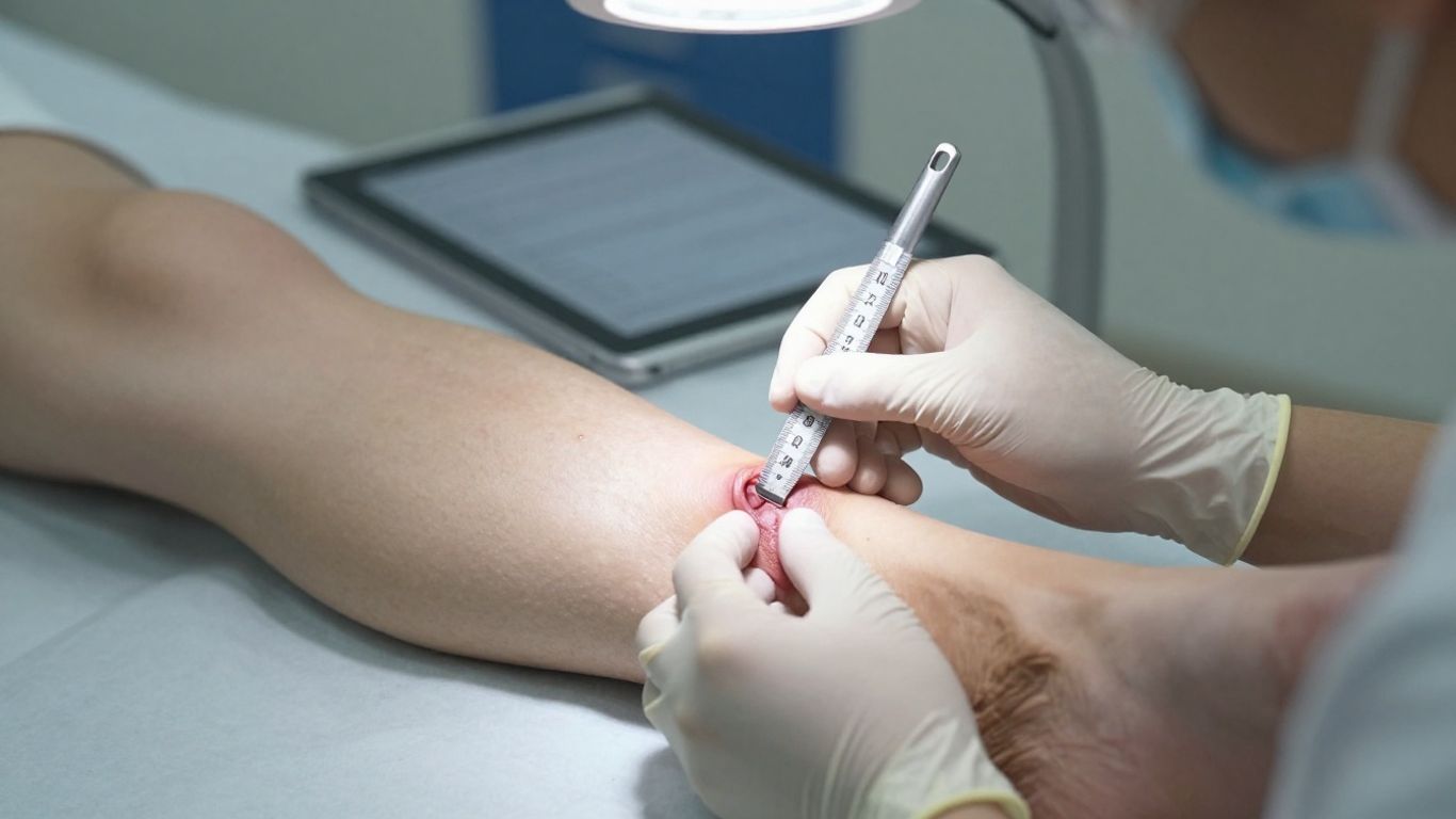
Evaluating wound size, depth, tissue, and drainage
Measure length, width, and, when appropriate, depth using a consistent method. Describe the wound bed, edges, surrounding skin, undermining, tunneling, tissue type, and the amount and character of exudate. Note whether the wound is covered by slough, eschar, granulation tissue, or epithelium.
Photographs can supplement—not replace—direct examination and measurements. Consistent positioning, lighting, and scale make serial images more useful, particularly when the change is gradual.
Identifying infection and other complications
Pain that is new or disproportionate, spreading redness, warmth, swelling, purulent drainage, malodor, friable tissue, or unexpected deterioration may raise concern for infection. Fever and systemic symptoms require prompt attention, but their absence does not exclude a serious local problem, especially in people with diabetes or impaired immunity.
Clinicians also look for undermining, necrosis, allergic contact dermatitis, maceration, bleeding, exposed structures, and signs of pressure or friction. These findings can change both the urgency and the treatment approach.
Assessing blood flow with ABI and vascular testing
An ankle-brachial index, or ABI, compares ankle and arm pressures as an initial assessment of lower-extremity perfusion. Results must be interpreted in context because arterial calcification can make vessels appear less compressible and may reduce the reliability of ABI alone.
Depending on the findings, the team may consider toe pressures, Doppler waveforms, skin perfusion measurements, or vascular imaging. An ischemic wound may not heal until impaired arterial supply is addressed, so repeated debridement without vascular review can be unsafe.
Reviewing diabetes, nutrition, medications, and mobility
A complete review includes glucose patterns, renal and cardiovascular disease, immune status, medications, nutritional intake, smoking, pain, sleep, mobility, and the ability to perform wound care at home. You may also need assessment of footwear, gait, transfer technique, or the support available from caregivers.
These details reveal practical barriers that a wound measurement cannot show. A technically appropriate plan still needs to be feasible, understood, and sustainable between visits.
Choosing the right wound care therapies
Therapy selection follows assessment and wound-bed preparation. The same wound may need different interventions at different stages, while two wounds with similar measurements may require different care because their causes differ. Clinicians weigh tissue viability, exudate, infection risk, perfusion, patient comfort, cost, and available support.
The aim is to create conditions in which the body can repair tissue. No advanced product substitutes for pressure relief, adequate blood flow, appropriate glucose management, or treatment of infection when those factors are present.
Debridement options for removing nonviable tissue
Debridement removes dead, damaged, or contaminated tissue that can obscure the wound bed and interfere with healing. Depending on urgency, tissue type, pain, perfusion, and setting, options may include sharp or surgical debridement, conservative sharp techniques, autolytic methods, enzymatic products, or mechanical approaches.
The choice should be individualized. Stable, dry eschar over an ischemic heel, for example, should not be treated in the same way as loose slough in a well-perfused wound.
Dressings that manage moisture and exudate
A dressing should maintain an appropriate moist environment while protecting surrounding skin and managing drainage. Foam, alginate, hydrofiber, absorbent contact layers, and other categories may be useful in different circumstances; the best choice depends on exudate, depth, tissue, frequency of change, and tolerance.
The surrounding skin deserves equal attention. Excess moisture can cause maceration, while an overly dry wound bed may slow cell movement. Reassessment is needed when drainage increases, decreases, or changes in character.
Negative pressure wound therapy (NPWT)
NPWT applies controlled suction through a sealed dressing to manage exudate and support wound-bed management in selected wounds. It may be considered for appropriately assessed surgical wounds, traumatic wounds, or other complex defects, but it is not suitable for every wound.
Before use, clinicians confirm adequate perfusion, control untreated infection when necessary, protect exposed structures, and select a seal and pressure setting appropriate to the patient. The dressing and tubing also require careful monitoring between changes.
Skin substitutes, grafts, and cellular therapies
Skin substitutes and grafts may be considered when a wound has a prepared, viable bed but is not closing as expected. Their use depends on wound etiology, perfusion, infection control, depth, available tissue, and the patient’s overall condition.
A biomaterial approach may also be discussed in selected cases. CholeDerm® is described by Alicorn Medical as a tissue-engineered wound care product derived from mammalian gall bladder extracellular matrix for acute and chronic wounds, surgical sites, and ulcers. Its use remains a clinical decision within an individualized plan, not a replacement for diagnosis or wound-bed preparation.
Advanced treatments for difficult-to-heal wounds
Difficult wounds are managed according to their underlying mechanism. Diabetes, venous hypertension, arterial insufficiency, pressure, neuropathy, edema, and infection each create different barriers to repair. A treatment plan becomes more coherent when those barriers are named explicitly.
For clinicians and patients, the practical question is not simply which product is advanced. It is whether the intervention addresses the specific reason the wound has stalled.
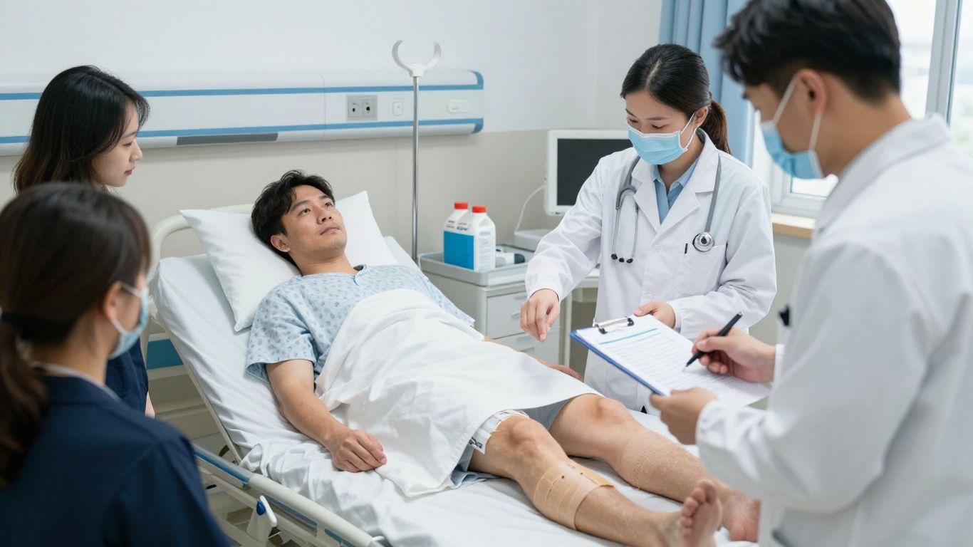
Care approaches for diabetic foot ulcers
Diabetic foot care commonly combines glucose management, regular inspection, debridement when appropriate, infection assessment, footwear review, and off-loading. Neuropathy can reduce protective sensation, so a small area of repetitive pressure may become a deep ulcer before it is noticed.
Inspect the entire foot, including between the toes and beneath pressure points. Management may require podiatry, vascular review, orthotics, diabetes care, and education for the patient and family.
Treatment strategies for venous leg ulcers
Venous leg ulcers are often associated with edema, venous hypertension, skin changes, and reduced calf-muscle pump activity. Compression can support edema control when arterial supply is adequate, but the method and pressure should be selected after vascular assessment.
Elevation, movement, appropriate dressings, skin protection, and treatment of venous disease may work together. Compression should not be applied casually when significant arterial insufficiency has not been excluded.
Managing pressure injuries and pressure relief
Pressure injuries develop when sustained pressure, shear, or friction compromises tissue. Treatment therefore includes repositioning, support surfaces, heel protection, moisture management, nutrition assessment, and attention to transfers and seating.
Relief must be continuous enough to matter. A wound can deteriorate if pressure is removed during a visit but returns for the rest of the day through bed position, wheelchair fit, or limited ability to move independently.
Addressing arterial and ischemic wounds
Arterial wounds may be painful, sharply demarcated, pale, punched out, or associated with cool skin and diminished pulses. They require careful perfusion assessment and timely vascular input, particularly when tissue loss is progressing or pain is severe at rest.
Avoid assuming that local wound treatment alone will restore healing. Revascularization decisions, medication review, smoking cessation, pain management, and protection from trauma may all be part of the pathway.
Managing wound infection and inflammation
Inflammation is a normal phase of repair, whereas infection is a clinical problem that can prevent progression and threaten surrounding tissue. Swab results alone do not determine the diagnosis because open wounds are commonly colonized by microorganisms. The examination, history, trajectory, and systemic condition must be considered together.
When infection is suspected, the plan may include source control, debridement, appropriate sampling, antimicrobial treatment, imaging, and escalation to hospital or specialist care. The urgency depends on depth, spread, perfusion, immune status, and the patient’s general condition.
Distinguishing colonization from clinical infection
Colonization means microorganisms are present without causing the clinical signs of infection. Infection becomes more likely when there is spreading erythema, increasing pain, warmth, swelling, purulence, tissue breakdown, malodor with deterioration, or delayed healing accompanied by other concerning findings.
Repeatedly treating a positive swab without clinical correlation can expose patients to unnecessary antimicrobials and miss the actual cause of delayed healing. Assessment should be repeated when the wound changes.
When topical or systemic antimicrobials may be used
Topical antimicrobials may have a limited role in selected local situations, while systemic antibiotics are generally reserved for clinically significant infection or deeper involvement as determined by a qualified clinician. Choice, dose, duration, and route depend on the suspected organism, tissue depth, kidney function, allergies, and local guidance.
Antimicrobials should support—not replace—drainage control, debridement, pressure relief, perfusion management, and appropriate dressing care. Unnecessary or prolonged use can create avoidable adverse effects and resistance concerns.
Recognizing cellulitis, osteomyelitis, and sepsis risks
Rapidly spreading redness, warmth, swelling, worsening pain, fever, chills, confusion, low blood pressure, or marked weakness requires urgent medical assessment. Osteomyelitis should be considered when bone is exposed or palpable, the wound is deep or recurrent, or healing remains poor despite appropriate care.
Do not wait for a routine dressing change when systemic illness or rapidly progressive tissue damage is possible. Emergency evaluation may be necessary.
Coordinating cultures, imaging, and specialist care
Cultures are most useful when collected appropriately from clinically relevant tissue after cleansing and, when indicated, debridement. Imaging may help evaluate a foreign body, gas, deep abscess, or bone involvement, while vascular studies clarify whether poor perfusion is contributing.
Clear communication among wound care, infectious disease, surgery, radiology, vascular services, and primary care reduces fragmented decisions. The result should be one shared plan with defined review points.
Supporting healing throughout the body
A wound is affected by the body’s broader physiology. Healing requires oxygen delivery, immune function, protein synthesis, energy, cell signaling, and the ability to protect the area from repeated injury. Advanced treatment therefore includes practical support for the conditions that make repair possible.
You should receive advice that is specific to your health status rather than a generic list of supplements or restrictions. Coordination with your medical team is especially important when kidney disease, heart failure, diabetes, or medication interactions are present.
Improving blood glucose control
Poorly controlled blood glucose can impair immune responses and tissue repair, while sudden changes in treatment can also create risk. Glucose targets and medication adjustments should be individualized by the clinician managing diabetes, with attention to nutrition, infection, renal function, and hypoglycemia.
Foot inspection and protective footwear remain important even when glucose control is improving. Better numbers do not remove existing neuropathy or pressure exposure overnight.
Addressing protein, calorie, vitamin, and mineral needs
Inadequate energy or protein intake can limit collagen formation and immune function. A nutrition assessment may consider weight change, appetite, swallowing, gastrointestinal symptoms, food access, renal status, and the demands of the wound.
Supplements should be guided by identified needs rather than taken indiscriminately. A dietitian can help match protein, calories, vitamins, and minerals to the patient’s condition and treatment goals.
Optimizing circulation, compression, and off-loading
Circulation must be assessed before compression or aggressive local treatment is selected. When appropriate, compression can help manage venous edema, while off-loading devices, footwear changes, cushions, and positioning reduce mechanical stress on vulnerable tissue.
These interventions work only when they are used consistently and fit the patient’s daily life. A device that cannot be worn safely or tolerated is unlikely to protect the wound.
Managing pain, smoking, edema, and underlying disease
Pain control can improve sleep, mobility, dressing tolerance, and participation in care. Smoking cessation, edema management, medication review, and treatment of cardiovascular, renal, inflammatory, or immune conditions may also influence recovery.
A broader review is particularly valuable when a wound repeatedly opens after closure. The recurring problem may be pressure, footwear, edema, vascular disease, or a systemic condition that was never fully addressed.
Monitoring recovery and adjusting the treatment plan
Healing is not judged by one encouraging photograph. Clinicians compare measurements, tissue quality, drainage, pain, surrounding skin, function, and the patient’s overall health across visits. The treatment plan should change when the evidence changes.
A scheduled review also gives you an opportunity to report practical difficulties, such as dressing failure, pain during off-loading, inability to reach the wound, or problems obtaining supplies. Those details often explain why a theoretically sound plan is not working in practice.
Measuring progress with consistent wound documentation
Use the same measurement approach whenever possible, recording dimensions, depth, tissue type, exudate, odor, periwound condition, pain, and relevant interventions. Document pressure relief, compression, glucose issues, vascular findings, and signs of infection alongside the wound itself.
A simple trend is more informative than isolated numbers. Serial documentation can show whether the wound is shrinking, filling with healthy tissue, becoming less exudative, or deteriorating despite treatment.
Signs that a treatment is working
Progress may include gradual reduction in area and depth, healthier granulation, advancing epithelial edges, less drainage, reduced odor, improved pain, and better function. The surrounding skin may also become less macerated or inflamed.
Improvement should be plausible for the wound’s cause and stage. A smaller wound is encouraging, but it does not prove that infection, ischemia, or pressure has been resolved.
When to change therapies or escalate care
Reconsider the plan when measurements plateau, the wound enlarges, drainage changes, pain increases, new necrosis appears, or the patient cannot follow the current regimen. Escalation may involve repeat vascular testing, imaging, specialist referral, a different dressing strategy, additional debridement, or hospital assessment.
Clinical evidence can also support continuing the current approach. The goal is not to change treatment frequently, but to avoid allowing an ineffective plan to continue without review.
Preventing recurrence after the wound closes
Once the surface closes, the tissue may remain fragile for months. Continue protection, moisturization of intact surrounding skin, prescribed compression or off-loading, gradual return to activity, and regular inspection. Keep follow-up appointments when the original cause remains active.
For deeper clinical background on tissue-derived biomaterials, the PrisTINE™ process is described as an enzyme-free, detergent-free approach intended to preserve natural tissue architecture and critical biomolecules. More broadly, Alicorn Medical develops biomaterial-based products from natural tissue, while evidence and product use should always be interpreted within appropriate clinical protocols.
Conclusion
Advanced wound care treatment works best when it is treated as a continuing clinical process rather than a single product choice. Careful assessment, cause-directed therapy, systemic support, and consistent follow-up give you the best opportunity for durable healing and lower recurrence risk.
Frequently Asked Questions
What is advanced wound care treatment?
It is a coordinated approach for complex, chronic, or non-healing wounds that combines wound-bed care with treatment of underlying problems such as infection, pressure, poor circulation, diabetes, or edema.
When should you see a wound care specialist?
Seek assessment when a wound is deep, worsening, recurrent, infected, associated with reduced circulation, or not showing meaningful improvement with appropriate initial care.
Can advanced wound care prevent amputation?
Timely assessment and treatment may reduce complications, but no approach can guarantee prevention. The result depends on factors including infection, blood flow, tissue loss, diabetes control, and how quickly care begins.
How is a diabetic foot ulcer treated?
Care may include off-loading, debridement when appropriate, infection assessment, glucose management, footwear review, vascular evaluation, and regular inspection by the patient and clinical team.
Are antibiotics always needed for an open wound?
No. Open wounds can be colonized without being clinically infected. Antibiotics are generally considered when examination and other findings support infection, and the choice should be made by a qualified clinician.
How long does a complex wound take to heal?
Healing time varies with wound depth, cause, blood flow, infection, nutrition, glucose control, pressure, and overall health. Consistent measurements are more useful than relying on a fixed timetable.
What can you do after a wound closes to prevent recurrence?
Continue skin checks, pressure relief, prescribed compression or off-loading, appropriate footwear, management of underlying disease, and follow-up for conditions that caused the original wound.