
Key Takeaways
Biomaterials can help you create a wound environment that supports moisture control, tissue protection, granulation, and repair, but they work best when matched to the wound’s cause and the patient’s wider clinical needs.
- Chronic wounds often remain trapped in inflammation rather than progressing through normal repair.
- Material choice should reflect exudate, tissue quality, wound depth, infection risk, and mechanical demands.
- ECM scaffolds provide structural and biological cues that may support cell migration and remodeling.
- Debridement, pressure relief, compression, vascular assessment, and infection control remain essential.
- Product selection should follow clinical evidence, regulatory requirements, wound-bed assessment, and ongoing monitoring.
Understanding chronic wounds and the role of biomaterials
A chronic wound does not progress through the normal stages of healing within the expected time, often because local and systemic factors keep interrupting repair. Diabetes, ischemia, neuropathy, pressure, venous disease, infection, and immobility can all contribute. If you are evaluating biomaterials for chronic wound treatment, begin with the underlying cause rather than treating the dressing as a standalone intervention. The material should support a broader plan that addresses perfusion, pressure, inflammation, contamination, and patient-specific risks.
How chronic wounds differ from acute wounds
An acute wound usually moves through hemostasis, inflammation, proliferation, and remodeling in a coordinated sequence. A chronic wound may remain in a prolonged inflammatory state, with delayed granulation, impaired epithelialization, recurrent infection, or persistent slough. Its appearance can also offer clues: venous ulcers often occur near the malleoli, arterial ulcers may be deeper and located on the heel or lateral foot, and neuropathic ulcers commonly develop at pressure points.
The distinction matters because a dressing cannot correct every cause of delayed healing. A pressure-related wound still needs pressure redistribution, while an ischemic wound requires vascular assessment. You should also consider whether the wound is infected, colonized, dry, highly exudative, undermined, or associated with exposed structures before choosing a biomaterial.
Why prolonged inflammation delays healing
Inflammation is necessary at the beginning of repair, but persistent inflammation can damage extracellular matrix, impair fibroblast activity, and interfere with angiogenesis. Excess matrix metalloproteinase activity may degrade matrix proteins and growth factors faster than the wound can replace them. Biofilm, repeated trauma, poor perfusion, and uncontrolled blood glucose can reinforce this cycle.
This is why a material that simply covers the wound may be insufficient. You may need to reduce nonviable tissue, manage bacterial burden, control exudate, and correct mechanical or vascular problems before a scaffold can function effectively. A useful overview of this relationship is available in this chronic wound healing editorial, which discusses disrupted inflammation, extracellular matrix damage, and stalled angiogenesis.
The importance of moisture balance, exudate control, and tissue protection
A moist, protected wound environment can support cell migration, while excessive fluid can macerate surrounding skin and dilute local signals. A dry wound may adhere to the dressing and suffer painful removal, whereas an overly occlusive approach can trap fluid and worsen local problems. Your choice should therefore account for the amount and character of exudate, the condition of the periwound skin, and how often the dressing can be changed safely.
Tissue protection also includes atraumatic removal, cushioning, thermal stability, and prevention of external contamination. These practical features affect comfort and adherence as much as the biomaterial’s biological properties. For a broader review of current approaches, see this discussion of current wound biomaterials.
How biomaterials support the wound-healing environment
Biomaterials can absorb fluid, form a protective gel, retain moisture, provide mechanical support, or act as a temporary matrix for host-cell interaction. Some are designed to deliver or preserve biological signals; others mainly manage the wound’s physical environment. The intended role should be clear before application.
A biomaterial is not a substitute for wound-bed preparation. Instead, it can support the transition toward granulation and epithelialization when the wound has been assessed and the major barriers to repair are being addressed. That distinction helps you interpret claims appropriately and avoid expecting one dressing to solve a multifactorial problem.
Key properties to evaluate in a wound biomaterial
When you compare biomaterials, look beyond the product category. Two materials described as scaffolds or hydrogels may differ substantially in source tissue, porosity, processing, degradation, fluid handling, and immune response. The most useful choice is the one whose properties fit the wound and the intended clinical workflow.
A practical evaluation also includes storage, handling, fixation, dressing-change requirements, and compatibility with adjunctive treatments. These details can determine whether a promising material is usable in a busy clinical setting.
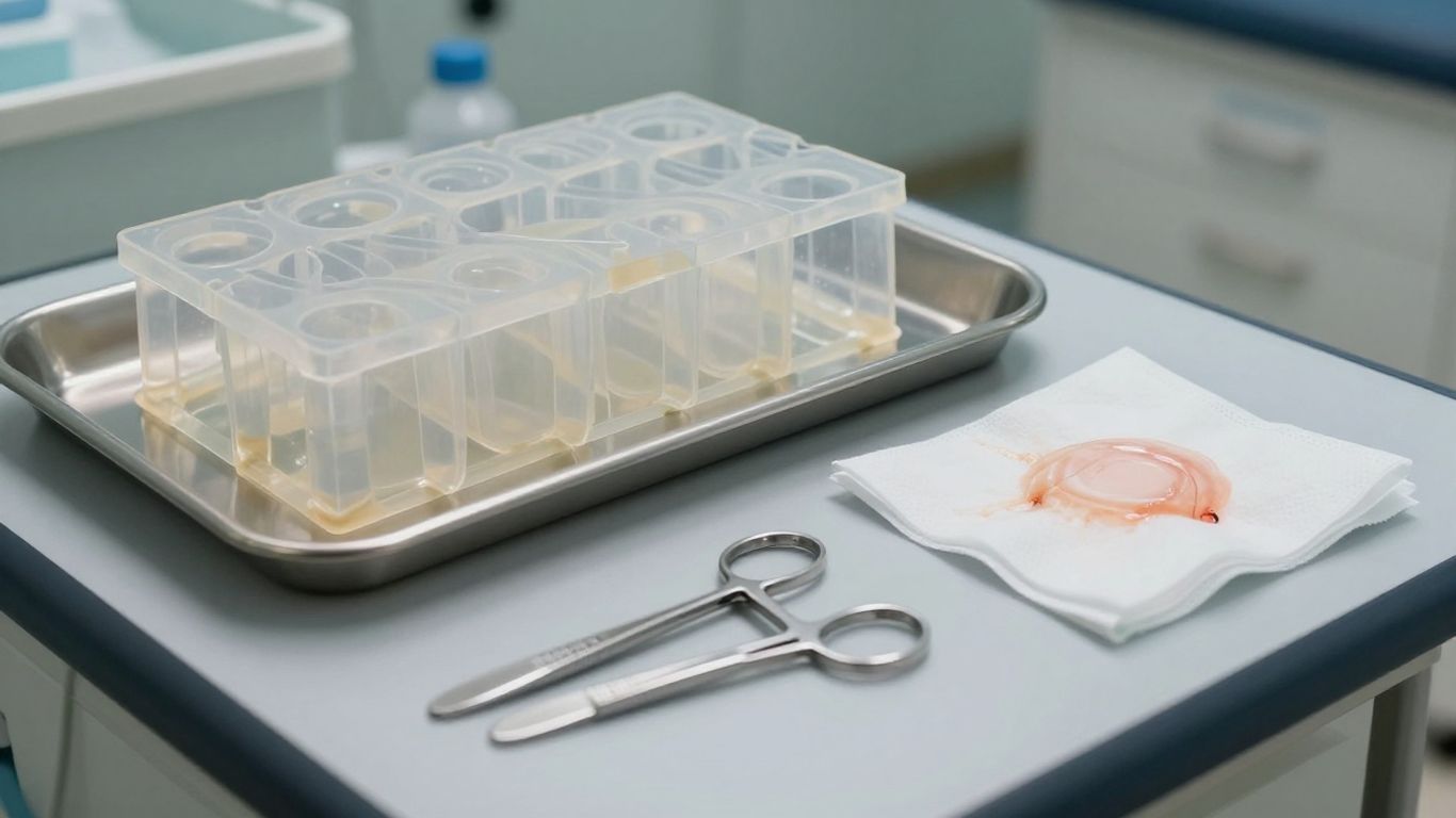
The image illustrates the basic clinical idea: a material must function at the interface between a complex wound bed and the surrounding care system. You still need product-specific instructions and appropriate clinical judgment before use.
Biocompatibility and immune response
Biocompatibility means more than the absence of an immediate visible reaction. You should consider cytotoxicity, inflammatory signaling, residual processing agents, source tissue, sterilization, and the likely host response over time. A biologic scaffold may interact with macrophages, fibroblasts, endothelial cells, and other host cells as it is incorporated or degraded.
Evidence from cell studies and animal models can help characterize these interactions, but it does not automatically predict human clinical outcomes. Look for clearly described methods, relevant endpoints, and evidence that distinguishes a controlled immune response from a claim of no immune response.
Porosity, structure, and cellular infiltration
The architecture of a material influences fluid movement, cell attachment, tissue infiltration, and mechanical behavior. A porous or fibrous structure may provide space for host cells and vascular ingrowth, while an overly dense material can limit penetration. Surface characteristics also affect how the material contacts the wound and whether it stays in place.
Natural ECM is more than collagen alone. It can include elastin, glycosaminoglycans, adhesion proteins, and other matrix-associated molecules that contribute to the local microenvironment. You should therefore ask whether processing preserves the structure and composition the product is intended to provide.
Degradation rate and tissue remodeling
A scaffold should remain present long enough to support the intended phase of repair, but it should not persist in a way that interferes with remodeling. Degradation depends on source, crosslinking, thickness, fluid exposure, enzymatic activity, and the condition of the wound. A material that degrades too quickly may lose structural value; one that remains too long may limit integration.
Controlled degradation is especially relevant when the goal is tissue replacement rather than temporary coverage. Research on ECM scaffolds has examined how stabilization and crosslinking can alter biodegradation, but the appropriate balance depends on the formulation and the clinical application.
Exudate management and moisture retention
Fluid handling is often the first property you notice at the bedside. Hydrofibers can form a gel, foams can absorb and cushion, and hydrogels can add moisture to a dry wound. You should match absorption capacity to exudate volume while protecting the periwound skin from leakage and maceration.
The material also needs to work with the secondary dressing and planned change interval. A highly absorptive primary layer may be useful in one wound but unnecessarily drying in another. Reassessment is essential because exudate commonly changes as inflammation and infection are controlled.
Antimicrobial function and biofilm considerations
Antimicrobial claims require careful interpretation. A material may act as a physical barrier, bind microorganisms, release an antimicrobial agent, or alter the wound environment. These mechanisms are not interchangeable, and in-vitro activity does not establish clinical effectiveness for every wound type.
Biofilm should be considered when a wound has recurrent slough, stalled progress, odor, or a slimy surface, although clinical assessment remains central. If an antimicrobial strategy is used, you should define its purpose, duration, tissue compatibility, and relationship to systemic infection management. Biomaterials with engineered antibacterial or antibiofilm functions remain an active area of research, as discussed in this overview of advanced wound biomaterials.
Common biomaterial-based dressings for chronic wounds
Each dressing class solves a different physical problem. You can think of the options as tools for fluid management, cushioning, matrix support, moisture adjustment, or contour adaptation. The right choice depends on wound depth, exudate, tissue quality, and the other interventions being used.
The categories below are not interchangeable prescriptions. They are a framework for discussing what each material is designed to do and when it may fit into a broader wound-care pathway.
Hydrofiber and gel-forming materials for exudative wounds
Hydrofiber materials are designed to absorb moderate to heavy exudate and form a gel that conforms to the wound bed. This can help limit lateral fluid spread and reduce the risk of maceration when the surrounding skin is protected appropriately. They may be considered for pressure ulcers, donor sites, burns, and other wounds with substantial drainage.
You should ensure that the dressing remains in contact with the wound without packing a cavity too tightly. A suitable secondary layer may be needed when exudate is heavy, and the wound should be reassessed rather than left under a fixed schedule when fluid levels change.
Foam dressings for absorption and cushioning
Foam dressings absorb excess exudate while providing cushioning and some thermal insulation. These features can be useful for pressure injuries, surgical wounds, graft sites, and wounds where protection from friction or impact matters. Their performance depends on thickness, absorbency, adhesive design, and whether the wound is producing more fluid than the foam can manage.
Foam is primarily a physical-management option. If the wound needs biological matrix support, antimicrobial treatment, debridement, or compression, those needs should be addressed separately rather than assumed to be covered by the foam.
Collagen-based products for slow-healing wounds
Collagen-based products are used in some slow-healing wounds because collagen can interact with the wound environment and provide a substrate associated with matrix formation. They may be considered for selected ulcers and surgical wounds when the wound bed has been prepared and excessive bioburden is being managed.
You should distinguish processed collagen products from intact or more complex ECM scaffolds. A collagen dressing may offer a focused matrix component, while a biologic scaffold can contain a broader structural and molecular profile. The product’s source, processing, and clinical evidence should guide that distinction.
Pastes, powders, and particulate materials for irregular defects
Particulate materials can be useful when a wound is deep, tunneled, undermined, or difficult to cover with a flat sheet. Pastes and powders may conform to irregular contours, absorb some exudate, and support contact with recessed areas. Some formulations also include antimicrobial or debriding functions, but those properties must be confirmed for the specific product.
Before using a particulate material, you should evaluate whether the cavity is clean, whether fluid can escape, and whether the material can be removed or monitored. A compact sequence can keep the application clinically grounded:
- Prepare the wound bed and remove nonviable tissue.
- Assess depth, tunneling, undermining, and exudate.
- Apply the material without overpacking the space.
- Add a compatible secondary dressing and document the response.
This approach keeps the material within a larger care plan. It also reduces the risk that a conformable product hides deterioration or prevents adequate reassessment.
Hydrogels and hybrid materials for moisture control
Hydrogels can donate moisture to dry wounds and may help soften adherent slough, depending on their formulation. Hybrid materials combine a hydrated polymer network with another structural or biological component, potentially allowing more deliberate control of handling, degradation, or signaling.
You should be cautious with hydrogels in heavily exudative wounds because added moisture can increase fluid burden. Emerging designs include materials intended to combine hydration with antimicrobial, antioxidant, or regenerative functions, but their clinical role depends on human evidence, product validation, and appropriate wound selection.
Extracellular matrix scaffolds and biologic dressings
ECM scaffolds are biologic materials intended to provide more than surface coverage. They can offer a three-dimensional architecture and matrix-associated molecules that interact with host cells during repair. Their value depends on the source tissue, decellularization or processing method, physical structure, immune response, and ability to integrate with the wound bed.
You should read biologic dressing claims carefully. Findings from a rabbit, rat, dog, or laboratory assay can explain mechanism and support further development, but they should not be presented as equivalent to controlled human clinical evidence.
How natural ECM supports cell migration and tissue repair
The native ECM provides a temporary environment in which cells attach, migrate, proliferate, and reorganize tissue. Collagen contributes structure, while elastin, glycosaminoglycans, adhesion proteins, and growth-factor-associated components can influence cell behavior. As the wound develops, the scaffold is expected to participate in a changing process rather than remain a permanent implant.
A porcine cholecyst-derived matrix has been investigated as a collagen-rich, fibrous or mesh-like scaffold with host-cell interaction and in-vivo degradation studied in preclinical work. Those findings support a scientific rationale for ECM research, but the model and formulation must remain explicit when discussing results.
Amniotic membranes and their signaling components
Amniotic membrane products are used in some wound-care settings because they may provide a biologic covering containing proteins, cytokines, and other signaling components. Their proposed roles include modulation of inflammation and support for regeneration in selected chronic wounds, burns, and postoperative wounds.
The clinical interpretation depends on preparation, preservation, thickness, viability or acellularity, storage, and regulatory status. You should not assume that every amniotic membrane product has the same biological activity or handling requirements.
Acellular dermal matrices from human, animal, and synthetic sources
Acellular dermal matrices may be derived from human or animal tissue, while synthetic scaffolds can be engineered to reproduce selected physical or chemical features. Human-derived matrices are used in some reconstructive applications, and animal-derived matrices may support dermal architecture, cellular infiltration, or graft-related procedures. Synthetic materials offer control over composition, porosity, and degradation, although they may not reproduce the full complexity of native ECM.
Source selection should follow the wound, intended use, available evidence, and product-specific information. You should also consider tissue origin, donor screening where relevant, crosslinking, antigen removal, sterilization, storage, and the requirements for fixation and coverage.
Why tissue origin and processing methods matter
Tissue origin affects collagen organization, elastin content, glycosaminoglycan composition, thickness, and the possible presence of cellular remnants. Processing then determines which of those features remain. Enzymes, detergents, chemical stabilization, decellularization methods, drying, and sterilization can all alter structure and biomolecule retention.
The research literature on cholecyst-derived ECM includes non-detergent and non-enzymatic recovery approaches, as well as separate work on stabilization and crosslinking. You should avoid reducing this complex process to vague claims such as “chemical-free,” and instead review the actual manufacturing and validation information.
Balancing biological activity, durability, and immunogenicity
A biologic scaffold must balance retained biological complexity with acceptable host response and sufficient handling strength. More processing may reduce some unwanted components but also remove useful matrix features. More stabilization may improve durability but change cellular access or degradation.
[CholeDerm®] is described in the supplied product material as a porcine, lyophilized, single-layer acellular dermal matrix containing collagen, elastin, glycosaminoglycans, and growth factors. The supplied research and product information should be read together, with commercial specifications and approved use kept distinct from preclinical findings.
Matching biomaterials to chronic wound type
Wound type provides a starting point, not a complete prescription. A diabetic foot ulcer may involve neuropathy, ischemia, pressure, and infection at the same time. A venous ulcer may require compression, while a pressure ulcer may not improve unless loading is reduced. Your biomaterial choice should follow the dominant barriers to healing and the wound’s current state.
Clinical history remains essential. Diabetes, peripheral vascular disease, neuropathy, trauma, immobility, malignancy, radiation, previous infection, medications, and comorbidities can all change the risk-benefit balance.
Biomaterial considerations for diabetic foot ulcers
For a diabetic foot ulcer, assess perfusion, sensation, pressure distribution, glycemic control, infection, depth, and exposed structures. A material that supports a moist environment may be useful, but it cannot compensate for ischemia or repeated mechanical loading. Offloading and vascular evaluation should proceed alongside wound-bed preparation.
ECM-based materials have been investigated in diabetic wound models, including a gelatin-modified cholecyst-derived scaffold studied in endothelial assays and a diabetic-rat full-thickness wound model. You should describe such findings as preclinical evidence from the evaluated formulation and model, not as a general human treatment guarantee.
Selecting materials for venous leg ulcers
Venous ulcers commonly occur around the gaiter area and may have irregular margins, granulation, slough, edema, and persistent exudate. Compression remains central when arterial supply is adequate and compression is clinically appropriate. A dressing or scaffold must fit beneath the compression system, manage fluid, and protect the periwound skin.
A biologic matrix may be considered when the wound has been prepared and remains stalled despite appropriate foundational care. Its use should not replace evaluation of edema, venous disease, infection, or adherence to compression.
Managing pressure ulcers and wounds associated with immobility
Pressure ulcers reflect sustained loading, shear, friction, moisture, and often reduced mobility or nutritional reserve. You should first address repositioning, support surfaces, seating, moisture management, nutrition, and pain. A biomaterial can protect or manage the wound, but ongoing pressure may prevent it from integrating or progressing.
For deeper wounds, assess undermining, tunneling, exposed bone or tendon, and the possibility of osteomyelitis. Absorbent or conformable materials may help with exudate, while a scaffold may be considered only after the wound bed and mechanical environment are suitable.
Addressing burns, surgical wounds, and traumatic tissue loss
Burns, surgical wounds, donor sites, and traumatic tissue loss vary widely in depth, contamination, perfusion, and need for reconstruction. A superficial wound may need protection and moisture control, while a full-thickness defect may require a dermal scaffold, graft support, flap, or staged reconstruction. The same material should not be assumed to fit every depth or anatomical site.
Product information for a specific biologic dressing should clarify its approved indications, preparation, fixation, secondary coverage, and relationship to grafting or surgery. Your plan should also account for pain, bleeding, exudate, and the need to inspect the wound regularly.
When ischemia, neuropathy, or infection changes the treatment plan
Ischemia can prevent oxygen and nutrient delivery, neuropathy can remove protective pain signals, and infection can accelerate tissue damage. These factors may change the sequence of care before any advanced biomaterial is applied. Vascular assessment, pressure relief, cleansing, debridement, cultures when clinically indicated, and systemic treatment may take priority.
A stalled wound should prompt reassessment rather than repeated application of the same dressing. If the wound worsens, develops spreading erythema, increasing pain, systemic symptoms, necrosis, or exposed deep structures, escalate evaluation promptly.
Combining biomaterials with advanced wound therapies
Biomaterials often work best as one part of a coordinated pathway. Negative pressure wound therapy, debridement, compression, offloading, irrigation, and infection control address different barriers. Combining them requires attention to sequencing, contact layers, fluid egress, fixation, and the risk of damaging the scaffold.
The aim is not to add every available technology. It is to select complementary interventions that support a measurable clinical goal, such as reducing exudate, preparing a wound bed, improving granulation, protecting a graft, or managing mechanical stress.
Using negative pressure wound therapy for exudate and granulation
Negative pressure wound therapy can help manage exudate, support wound contraction, and create mechanical conditions associated with granulation in selected wounds. You should first ensure that the wound is adequately debrided, that bleeding is controlled, and that exposed structures are protected according to the treatment protocol.
The foam, seal, pressure setting, and change interval all matter. NPWT is not appropriate for every wound, and contraindications and precautions must be reviewed before use. Its role should be judged by wound progress, fluid control, tissue quality, and patient tolerance.
Combining ECM scaffolds with negative pressure wound therapy
An ECM scaffold can provide a biological interface, while NPWT provides mechanical support and fluid management. The combination may be considered for large, deep, or highly exudative wounds when the clinical team can protect the scaffold and maintain adequate fluid egress.
[CholeDerm®] product guidance describes hydration, a non-adherent contact layer, and placement of NPWT foam and dressing as part of a combined workflow. The supplied material also describes fenestration or meshing for high-exudate wounds to permit fluid movement. Follow current approved instructions and clinician training rather than generalizing this workflow to unrelated products.
When irrigation or topical oxygen may be considered
Irrigation can help remove debris and dilute surface contamination during wound cleansing, provided the solution and pressure are appropriate for the tissue. It should not substitute for debridement when nonviable tissue is present. Topical oxygen may be considered in selected settings, but its role depends on the wound, equipment, protocol, and quality of supporting evidence.
These approaches should be integrated with assessment of perfusion and infection. A technology that changes the local environment cannot overcome severe arterial insufficiency or uncontrolled systemic infection on its own.
Integrating debridement, offloading, compression, and infection control
A biomaterial is most likely to help when the wound bed is viable and the main mechanical and infectious barriers are being addressed. In practice, you may need to coordinate several steps:
- Debride nonviable tissue and cleanse the wound.
- Reduce pressure, shear, and repetitive trauma.
- Use compression when venous disease is present and arterial supply permits.
- Control local or systemic infection according to clinical findings.
After these measures, select a material that matches exudate and tissue requirements. Document wound dimensions, tissue composition, drainage, pain, periwound condition, and the response at each review so that the plan can change when the wound changes.
Using antimicrobial strategies without encouraging resistance
Antimicrobial treatment should have a defined indication and duration. Overuse of antibiotics can contribute to resistance, while indiscriminate antiseptic exposure may injure viable tissue. Physical approaches that bind or remove microorganisms, antimicrobial coatings, and carefully selected chemical agents each have different mechanisms and evidence requirements.
You should distinguish local bioburden management from treatment of invasive infection. If systemic signs or spreading infection are present, a dressing strategy alone is not adequate. Repeated assessment helps determine whether an antimicrobial intervention is reducing the problem or merely masking a stalled wound.
Evaluating evidence and implementing biomaterials safely
Evidence for biomaterials exists across a spectrum, from material characterization and cell assays to animal studies, case reports, comparative trials, and systematic reviews. These layers answer different questions. A laboratory result may explain how a scaffold interacts with cells, while a clinical study is needed to evaluate safety, usability, healing, and patient-relevant outcomes.
Implementation also depends on training, storage, preparation, fixation, documentation, and access to multidisciplinary support. A clinically promising material can fail in practice if the surrounding pathway is inconsistent.
Distinguishing preclinical findings from human clinical evidence
Preclinical studies can examine angiogenesis, granulation, epithelialization, inflammation, degradation, and tissue integration under controlled conditions. They are valuable for mechanism and safety development, but animal wound models do not reproduce every feature of a human chronic wound. You should preserve the model, formulation, comparator, and endpoint when describing the result.
Human evidence should be assessed for study design, sample size, wound type, follow-up, comparator care, adverse events, and the definition of healing. A single case or uncontrolled series may suggest feasibility, but it cannot establish that a product will produce the same result in every patient.
Assessing wound-bed readiness before application
Before applying a scaffold or biologic dressing, confirm that the wound has been assessed for etiology, perfusion, infection, depth, exudate, and tissue viability. Remove or manage nonviable tissue as clinically indicated, control bleeding, and protect exposed structures. The wound should be sufficiently clean and viable for the intended material to contact the target tissue.
Readiness is not a permanent state. A wound can become more exudative, contaminated, ischemic, or painful after application. Your protocol should specify when to inspect the wound and what findings require removal, escalation, or a change in treatment.
Monitoring granulation, exudate, epithelialization, and adverse reactions
Track measurable changes rather than relying only on visual impressions. Record wound area and depth, granulation quality, slough or necrosis, drainage amount, odor, epithelial advancement, pain, periwound changes, and signs of inflammation or infection. Photographs can support comparison when obtained consistently and with appropriate consent.
You should also monitor material-specific reactions, including persistent erythema, excessive pain, bleeding, maceration, displacement, or delayed deterioration. If progress stalls, revisit the diagnosis and the care pathway instead of simply extending the same application schedule.
Considering regulatory status, handling, and product validation
Regulatory classification, approved indications, storage conditions, shelf life, sterility or sterilization validation, packaging integrity, and preparation instructions are practical safety considerations. Research publications cannot substitute for current product documentation. You should confirm that the material is appropriate for the intended wound and that the clinical team can handle it as directed.
Validation should cover more than biological plausibility. It may include manufacturing consistency, physical properties, biocompatibility testing, packaging, transport, and instructions for use. These details support reliable implementation and help separate a research formulation from a commercially validated medical product.
Building a patient-specific treatment pathway with multidisciplinary care
Chronic wound care often involves wound specialists, nurses, surgeons, podiatrists, vascular clinicians, infectious disease professionals, nutrition teams, physiotherapists, and the patient or caregiver. Each contributor may address a different barrier to healing. Shared documentation reduces duplication and makes it easier to identify why a wound is improving or stalling.
[Alicorn Medical] develops biomaterial-based medical products from natural tissue, with research focused on tissue-engineered wound-care solutions and clinician and academic collaboration. Any product discussion should remain aligned with current approved information, while the patient-specific plan should remain grounded in assessment, evidence, and informed consent.
Conclusion
Biomaterials can give you useful ways to manage moisture, exudate, tissue protection, matrix support, and biological signaling in chronic wounds, but their value depends on appropriate wound selection and coordinated care. When you combine careful assessment with debridement, pressure or compression management, infection control, monitoring, and realistic interpretation of evidence, advanced materials become part of a safer and more purposeful treatment pathway.
Frequently Asked Questions
What are biomaterials for chronic wound treatment?
They are natural, synthetic, or hybrid materials used to protect a wound, manage moisture and exudate, provide structural support, interact with host tissue, or deliver specific biological or antimicrobial functions.
Can a biomaterial heal a chronic wound by itself?
Usually not. Chronic wounds often involve pressure, ischemia, neuropathy, diabetes, infection, venous disease, or other systemic factors that must be assessed and treated alongside the selected material.
How do you choose a biomaterial for a chronic wound?
Consider the wound’s cause, depth, tissue quality, exudate, infection risk, periwound condition, mechanical demands, and the evidence and approved use for the specific product.
What is the difference between a collagen dressing and an ECM scaffold?
A collagen dressing focuses primarily on collagen as a matrix component, while an ECM scaffold may preserve a broader three-dimensional structure and several matrix-associated molecules. Processing and product-specific evidence determine the practical difference.
Are ECM scaffolds suitable for diabetic foot ulcers?
They may be considered in selected cases, but only after assessment of perfusion, infection, neuropathy, pressure, glycemic factors, and wound-bed readiness. Offloading and vascular care remain essential.
Can biomaterials be used with negative pressure wound therapy?
Some materials may be combined with negative pressure therapy under appropriate protocols. The clinician must consider fluid egress, contact layers, fixation, pressure settings, exposed structures, and the product’s instructions for use.
How should progress be monitored after applying a biomaterial?
Monitor wound dimensions, granulation, epithelialization, exudate, odor, pain, periwound condition, infection signs, material displacement, and adverse reactions at defined review points.