
Key Takeaways
CholeDerm wound dressing is a lyophilised, unmeshed acellular dermal matrix intended for use as part of a structured wound-care plan. Its role, application, and follow-up depend on wound-bed quality, perfusion, infection control, exudate, and the patient’s underlying condition.
- It is a single-layer, porcine-derived extracellular matrix scaffold.
- The matrix contains collagen, elastin, glycosaminoglycans, and growth factors.
- It may be considered for several partial- and full-thickness wounds, ulcers, burns, and surgical wounds.
- Preparation, hydration, fixation, protection, and moisture control are central to application.
- NPWT, compression, offloading, and treatment of the underlying cause may remain necessary.
What CholeDerm wound dressing is
CholeDerm wound dressing is described in the product specifications as a lyophilised, unmeshed, single-layer acellular dermal matrix of porcine origin. You can think of it as a biologic scaffold placed directly against a prepared wound bed, rather than as a conventional absorbent cover. The clinical decision still rests on assessment, wound-bed preparation, and the wider plan of care.
Acellular dermal matrix and extracellular matrix structure
An acellular dermal matrix is tissue from which cellular components have been removed while the supporting extracellular matrix is retained. That structure provides a physical interface for contact with the wound bed and a framework through which repair processes may proceed. The matrix is less than 1 mm thick, specified as approximately 100 μm.
Porcine source and biological composition
The documented source is porcine tissue, specifically a gall-bladder-derived extracellular matrix. This matters clinically because you should record the animal origin during product selection and patient screening. The material is supplied as a sterile, lyophilised sheet for single use.
Collagen, elastin, GAGs, and growth factors
The listed base material includes collagen, elastin, glycosaminoglycans, and growth factors. These components are naturally associated with dermal structure, hydration, cell interaction, and repair signalling. The source material also describes preservation of native extracellular-matrix components through a non-enzymatic, non-detergent process.
How the scaffold supports wound healing
The scaffold is intended to provide biological and structural support while the wound progresses through granulation and epithelialization. Preclinical material describes findings including angiogenesis, tissue remodelling, organized granulation, and early epithelialization across several wound models. Those findings should guide understanding of the mechanism, not replace patient-specific clinical judgment.
Regulatory, manufacturing, and sterilization considerations
The product specifications identify CholeDerm as a CDSCO-approved Class D device, while the supplied material describes a lyophilised and sterile sheet. You should verify the current package label, instructions for use, lot information, and institutional requirements before treatment. The associated SCTIMST research background also places the scaffold in the context of indigenous extracellular-matrix development and clinical translation.
Which wounds may benefit from CholeDerm
The potential wound types are broad, but that does not mean every wound is an automatic candidate. You should first establish whether the wound is adequately perfused, appropriately debrided, and free of uncontrolled infection. CholeDerm is best considered as one part of a plan that also addresses pressure, venous disease, diabetes, neuropathy, or vascular compromise.
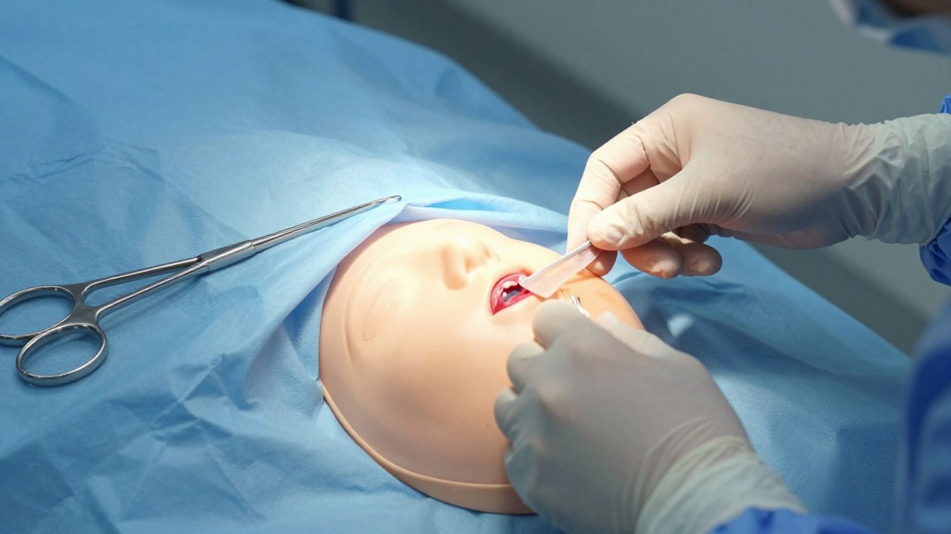
Partial- and full-thickness wounds
The stated indications include both partial-thickness and full-thickness wounds. These wounds differ in depth, tissue loss, exudate, and the amount of dermal support required, so the same application strategy may not suit both. Measure the wound and assess the visible tissue before selecting a matrix size or deciding whether additional intervention is needed.
Diabetic, venous, pressure, and vascular ulcers
The indication list includes diabetic ulcers, pressure ulcers, venous ulcers, and chronic vascular ulcers. In these cases, local coverage alone cannot correct ischemia, venous hypertension, repetitive pressure, or neuropathy. Compression, offloading, glucose management, vascular assessment, and pressure relief may therefore be essential companions to the dressing.
Surgical wounds, dehiscence, and donor sites
Surgical wounds, wound dehiscence, donor or graft wounds, post-Mohs wounds, post-laser wounds, and podiatric wounds appear within the documented use categories. You should clarify whether the wound requires drainage, re-closure, grafting, or treatment of a deeper complication before applying a biologic matrix. The matrix may be considered as an adjunct when the wound is suitable for local wound management.
Burns, trauma wounds, and skin tears
The listed categories include second-degree burns, trauma wounds, abrasions, lacerations, and skin tears. Fragile tissue needs gentle cleansing, atraumatic contact layers, and careful adhesive selection. Burn depth, contamination, pain, fluid loss, and the need for specialist assessment should be evaluated independently of dressing choice.
Complex, tunneled, and undermined wounds
Tunneled and undermined wounds are included in the product’s indication list, as are draining wounds. These wounds require careful probing, documentation of depth and direction, and attention to dead space and exudate. A sheet should not be forced into a cavity; packing or a different configuration may be needed according to the clinical assessment.
When to consider CholeDerm in a treatment plan
You might consider an acellular matrix when a wound remains stalled despite appropriate standard care, or when it could serve as a bridge to a later procedure. The decision should be based on wound trajectory rather than appearance alone. A clear plan for debridement, moisture management, reassessment, and treatment of the cause makes the intervention more meaningful.
Non-healing wounds despite standard care
Chronic wounds that show little granulation despite appropriate cleansing, pressure relief, compression, or other indicated care may warrant review by a wound specialist. The supplied clinical material includes case reports and preliminary observations, but individual outcomes should not be treated as guarantees. Before escalation, revisit perfusion, infection, adherence, nutrition, glycaemic control, and mechanical stress.
Preparing a wound bed for grafting or reconstruction
An extracellular matrix may be considered as a bridge while a wound bed becomes more suitable for grafting or reconstruction. The source material describes its use as an adjunct to definitive procedures and as support before delayed surgery. You should define the intended endpoint: granulation, epithelialization, reduction in wound size, or readiness for another intervention.
Supporting healing when surgery carries higher risk
For patients with substantial comorbidity, radiated tissue, diabetes, or vascular compromise, surgery may carry increased risk. The clinical material describes the device as an alternative when surgery is undesirable or contraindicated, but that decision belongs within specialist assessment. A conservative pathway should still include escalation criteria if the wound deteriorates or fails to progress.
Combining ECM support with compression and offloading
An extracellular matrix does not remove the need to control the forces that caused the wound. Venous ulcers may still require compression when appropriate, while plantar or pressure-related wounds may need offloading. In practice, the dressing works within a larger plan that protects the wound from recurrent trauma and manages exudate.
Assessing perfusion, infection, and underlying causes
Before application, assess pulses or other relevant indicators of perfusion, wound dimensions, tissue quality, exudate, odour, pain, and surrounding skin. Address infection, necrosis, biofilm concerns, pressure, neuropathy, and vascular disease as indicated. The clinical development and approval context can provide background, but it does not replace bedside assessment or local protocols.
How to apply CholeDerm wound dressing
Application follows a practical sequence: prepare the bed, size the sheet, place it in contact with viable tissue, hydrate it, secure it, and protect it. The exact technique should follow the current instructions for use and your organisation’s policy. The aim is to preserve contact with the wound while maintaining a manageable moisture environment.
Debriding and preparing the wound bed
Thorough debridement is described as the first step, with removal of non-viable tissue and control of bleeding and excess exudate. Cleanse the wound according to the clinical situation and confirm that the remaining bed is viable. If the bed is necrotic, heavily contaminated, or actively infected, resolve those issues before proceeding whenever clinically appropriate.
Trimming and placing the matrix
Trim the sheet to the wound dimensions, allowing it to extend slightly beyond the wound margins where appropriate. Place it directly against the prepared tissue, avoiding folds and unsupported bridging. Handle the matrix with sterile technique and minimise manipulation once it is positioned.
Hydrating with sterile saline
The supplied workflow specifies hydration with sterile saline after placement. Hydration helps the sheet conform to the wound surface and supports close contact. Use enough fluid for the intended configuration without creating avoidable pooling beneath the dressing.
Fixing the dressing and protecting the wound
The matrix may be secured with sutures, staples, or sterile adhesive strips according to the site, tissue quality, and local practice. A porous, non-adherent primary protective dressing is then placed over it to reduce disruption during subsequent changes. Avoid fixation methods that create unnecessary trauma to fragile surrounding skin.
Managing moisture with secondary dressings
Secondary dressing selection should follow the amount of exudate and the condition of the wound. The practical options can be organised as follows:
| Wound situation | Secondary dressing approach | Clinical aim |
|---|---|---|
| Dry wound | Moisture-retentive layer | Prevent desiccation |
| Low exudate | Protective, non-adherent cover | Preserve contact and comfort |
| Moderate to high exudate | Absorbent secondary layers | Control fluid and protect periwound skin |
| Large or deep wound | Layered protection with close review | Manage dead space and exudate |
This framework is only a starting point. Reassess the periwound skin and the wound surface at each change, then adjust absorbency or moisture retention as the wound evolves.
Using CholeDerm with NPWT
Negative pressure wound therapy can be used as an adjunct in selected large, deep, high-exudate, or high-risk wounds. The supplied workflow places the hydrated matrix against the wound, followed by a non-adherent contact layer, NPWT foam, and a sealed dressing. You should use the pressure settings, interface, and change schedule prescribed for the patient and wound.
Why ECM and negative pressure can complement each other
The source material describes ECM as a biological interface and NPWT as a form of mechanical support. NPWT may assist with exudate control, wound contraction, and perfusion, while the matrix provides the extracellular-matrix scaffold. These roles are complementary, but they do not eliminate the need to treat infection, pressure, ischemia, or other causes.
Applying the matrix and non-adherent contact layer
Apply and hydrate the matrix directly on the prepared wound, then cover it with a non-adherent contact layer. This interface helps protect the scaffold from direct adherence to the foam and can support less traumatic dressing changes. Ensure that the contact layer does not obstruct intended fluid movement or leave untreated wound areas exposed.
Managing exudate in large or deep wounds
High exudate can cause leakage, maceration, seal failure, and discomfort. Choose the foam and surrounding dressing configuration according to the wound size and fluid burden, and inspect the seal and periwound skin at scheduled reviews. A sudden change in output, odour, pain, or tissue appearance should prompt reassessment rather than simply increasing suction.
When fenestration may support fluid egress
For high-exudate wounds, the supplied guidance allows fenestration or meshing of the matrix with a scalpel to support fluid egress under NPWT. This is a procedural decision and should be performed only by a trained clinician using sterile technique. The modification should preserve meaningful wound coverage while reducing the chance of fluid collecting beneath the matrix.
Patient and wound types suited to combination therapy
The documented examples include large or deep wounds, diabetic-foot wounds, post-surgical wounds, complex wounds, and wounds with high exudate. Suitability depends on the wound bed, perfusion, infection status, patient tolerance, and ability to maintain a seal. NPWT should be stopped or revised if the wound shows deterioration or if therapy is no longer clinically appropriate.
Selecting sizes, packaging, and storage options
Product selection should match the wound area while allowing practical placement and fixation. The specification lists five sheet dimensions and two packaging configurations. Before opening a pack, confirm the product identity, size, lot details, package integrity, and storage conditions.
Available CholeDerm sheet dimensions
The listed sheet dimensions range from 3.6 × 3.0 cm to 7.6 × 4.6 cm. The available models are shown below for quick comparison.
| Model | Sheet size | Area |
|---|---|---|
| CD-P3630-1S | 3.6 × 3.0 cm | 10.8 cm² |
| CD-P4636-1S | 4.6 × 3.6 cm | 16.56 cm² |
| CD-P5640-1S | 5.6 × 4.0 cm | 22.4 cm² |
| CD-P6646-1S | 6.6 × 4.6 cm | 30.36 cm² |
| CD-P7646-1S | 7.6 × 4.6 cm | 34.96 cm² |
These areas are product specifications, not a substitute for measuring the wound after preparation. Wound shape, margins, tunnels, and the need for overlap can affect the practical choice.
Choosing a sheet based on wound area
Measure length and width after cleansing and debridement, and consider depth and irregularity separately. A sheet that is too small may leave exposed wound margins, while excessive trimming can create unnecessary waste. Document the selected size and the reason for selection in the clinical record.
Single-sheet and five-sheet packaging
The specifications list one-sheet and five-sheet packs. A single-sheet pack may suit an isolated application, while a five-sheet pack may be practical when several procedures or staged changes are anticipated. Follow facility policy for traceability and do not use a pack if its sterile barrier is compromised.
Room-temperature storage requirements
Storage is specified at room temperature up to 25 °C. Keep the product in its original packaging, protected from conditions that could compromise sterility or material integrity. If storage conditions have been exceeded, seek guidance before use rather than relying on visual appearance alone.
Shelf life and handling checks before use
The listed shelf life is two years, subject to the labelled expiry date and storage conditions. Before opening, check the expiry, package seal, label, and any visible damage. Once opened, use the sheet according to the instructions for use and discard any unused material when required by the product label or local policy.
Safety, contraindications, and clinical monitoring
Safety depends on patient selection, sterile handling, appropriate wound preparation, and timely review. You should document baseline wound measurements, tissue type, exudate, pain, surrounding skin, and any signs of infection. Monitoring should continue after application rather than ending when the dressing is secured.
Screening for porcine allergy
Because the matrix is porcine-derived, ask about known porcine allergy or sensitivity and document the result before use. Consider the patient’s cultural, religious, and personal preferences as part of informed consent and product selection. If the history is uncertain or concerning, pause and seek appropriate clinical advice.
Treating infection before application
Uncontrolled infection, spreading cellulitis, systemic illness, or untreated necrotic tissue requires priority management. Look for increasing pain, warmth, erythema, purulence, malodour, fever, or other systemic signs. A biologic scaffold should not be used to conceal a wound that needs urgent antimicrobial, surgical, or vascular treatment.
Monitoring granulation, epithelialization, and exudate
At follow-up, record wound dimensions, granulation, epithelial advancement, tissue quality, exudate amount and character, and periwound condition. Healthy progress may include a cleaner wound bed, viable granulation, reduced exudate, and advancing epithelialization. Photographs and consistent measurements can make gradual change easier to judge.
Recognizing signs of deterioration or complications
Escalate if the wound enlarges, pain increases unexpectedly, exudate becomes purulent or foul-smelling, bleeding persists, or surrounding redness spreads. Also assess for maceration, allergic reaction, dressing displacement, seal failure with NPWT, or signs of deeper infection. A change in the wound trajectory matters more than a single isolated measurement.
Reviewing dressing changes as the wound evolves
The dressing plan should change as exudate falls, granulation develops, and epithelialization progresses. Reconsider the secondary layer, fixation method, visit interval, compression, offloading, or NPWT configuration at each review. The advanced wound care overview offers broader product context, while bedside decisions should remain guided by the patient’s response and current clinical evidence.
Conclusion
CholeDerm wound dressing is most appropriately viewed as a porcine-derived extracellular-matrix scaffold used within a complete wound-care plan. Careful assessment, wound-bed preparation, hydration, fixation, moisture control, and follow-up determine whether the application is clinically appropriate. When needed, compression, offloading, NPWT, infection management, and treatment of underlying disease remain central to the patient’s progress.
Frequently Asked Questions
What is an acellular dermal matrix?
An acellular dermal matrix is biologic tissue processed to remove cellular components while retaining extracellular-matrix structures that can provide a scaffold at the wound interface.
Which wounds may require an advanced dressing?
Chronic ulcers, selected burns, surgical wounds, dehiscence, donor sites, trauma wounds, and other slow-healing wounds may require advanced dressings after assessment of depth, perfusion, infection, and exudate.
Why is wound-bed preparation necessary?
Debridement and cleansing remove non-viable tissue, debris, and excess contamination so that the selected dressing can contact a cleaner, viable wound bed.
How do you manage heavy wound exudate?
Use an absorbent secondary dressing, protect the periwound skin, review the underlying cause, and consider specialist strategies such as NPWT when clinically indicated.
When is compression used in wound care?
Compression is commonly considered for venous disease when arterial supply and other contraindications have been assessed. It should be prescribed and applied appropriately for the patient.
What signs suggest that a wound is worsening?
Increasing pain, spreading redness, warmth, purulent or foul-smelling drainage, enlarging dimensions, tissue necrosis, fever, or systemic illness should prompt timely clinical review.
How often should a wound dressing plan be reviewed?
Review timing depends on the wound, exudate, dressing, and patient risk. Reassess whenever the wound changes, the dressing fails, or new symptoms appear, and update the plan as healing progresses.