
Key Takeaways
Enzyme free tissue decellularization uses physical and osmotic steps to remove cellular material while seeking to preserve the native extracellular matrix.
- The process is tissue-specific; cellularity, thickness, and architecture shape the protocol.
- Mechanical delamination, freeze–thaw treatment, osmotic exposure, and rinsing can reduce reliance on enzymes and detergents.
- Preserving collagen, glycosaminoglycans, adhesion proteins, and growth factors may support tissue remodeling.
- Histology, DNA testing, protein profiling, mechanical assessment, and sterility testing are all needed to judge quality.
- A successful scaffold balances low residual cellular content with structural and biological preservation.
What enzyme free tissue decellularization means
When you work with a biological tissue, decellularization means removing cells and cellular remnants while retaining the extracellular matrix, or ECM. The ECM supplies structural organization and can also carry biochemical cues that influence how new cells attach, migrate, and remodel tissue. In enzyme free tissue decellularization, the process is designed around physical, osmotic, and washing steps rather than added proteolytic or nucleic-acid-digesting enzymes. The exact balance depends on the source tissue and the intended scaffold.
Definition and core objectives
The central objective is selective removal, not simply aggressive disruption. You want to reduce nuclei, DNA, membrane fragments, and other immunologically relevant cellular material without unnecessarily removing collagen, elastin, glycosaminoglycans, adhesion proteins, or soluble matrix-associated signals. A useful scaffold therefore reflects two outcomes at once: adequate decellularization and preservation of an organized ECM. The decellularization process is best understood as a tissue-engineering strategy rather than a single universal recipe.
How it differs from enzyme- and detergent-based methods
Enzyme- and detergent-based protocols may use lytic agents, surfactants, nucleases, proteases, or several combinations in sequence. An enzyme free approach instead relies on methods such as mechanical separation, freeze–thaw disruption, osmotic treatment, and repeated rinsing. This can reduce exposure to processing reagents, although it does not remove the need for careful validation. Physical methods can be effective in a suitable tissue, but the outcome still depends on geometry, cellular density, temperature control, and washing adequacy.
Why preserving the extracellular matrix matters
The ECM is more than a passive mesh. Its collagen network provides mechanical support, while proteoglycans and glycosaminoglycans influence hydration and local signaling. Adhesion proteins can help guide cell attachment and migration, and matrix-associated growth factors may contribute to the biological environment around a wound or engineered construct. For that reason, matrix preservation matters as much as visible cell removal when you assess a scaffold.
Common misconceptions about “enzyme free” processing
“Enzyme free” does not mean chemical-free, sterile by default, or automatically complete. It describes the absence of added enzymes in a defined processing workflow; osmotic solutions, buffers, temperature changes, and sterilization may still be used. It also does not guarantee that every tissue can be fully decellularized through physical steps alone. You still need tissue-specific criteria, validated operating conditions, and independent testing to establish safety and performance.
Why choose an enzyme free decellularization strategy
The appeal of an enzyme free decellularization strategy is its focus on limiting unnecessary alteration of the matrix. If the starting tissue is relatively thin and less cellular, physical handling may remove cellular layers without the long sequence of reagents often associated with more complex protocols. That can simplify process control, but it does not eliminate technical risk. Your goal remains a measured compromise between cellular clearance, biomolecule retention, structural fidelity, and sterility.
Protecting collagen and native tissue architecture
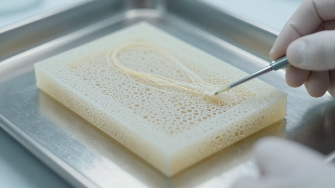
Collagen fibers form much of the load-bearing framework in many soft tissues. Harsh or prolonged processing can change fiber organization, swelling behavior, porosity, and mechanical response. A physical workflow may help you retain more of the starting architecture when the tissue’s anatomy permits gentle layer removal. The result should be checked rather than assumed through microscopy, morphology, and mechanical testing.
Retaining growth factors, glycosaminoglycans, and adhesion proteins
Biological activity can reside in several matrix components, not only in collagen. Glycosaminoglycans contribute to hydration, while proteins such as fibronectin and other adhesion molecules can influence how host or seeded cells interact with a scaffold. Growth-factor activity may also be affected by processing conditions. For a wound-oriented matrix, retention of these components is a practical reason to minimize unnecessary chemical exposure, while recognizing that preservation must be demonstrated analytically.
Reducing chemical residues and processing complexity
A shorter workflow can reduce the number of process variables you need to control and may limit concerns about residual detergents, enzymes, solvents, or cross-linking agents. It may also make rinsing and batch documentation more straightforward. However, fewer steps are not automatically better: every omitted step must be matched by adequate evidence that cellular debris, contamination, and unwanted residues have been addressed.
Balancing cellular removal with matrix preservation
You can think of protocol design as a series of trade-offs. More aggressive disruption may improve clearance but damage the ECM; gentler treatment may preserve structure but leave residual material. The most appropriate endpoint is therefore not maximum processing intensity, but a validated window in which residual cellular content is acceptably low and the scaffold remains biologically and mechanically useful.
Tissue selection and preparation
The starting tissue often determines whether an enzyme free workflow is practical. You should consider cellular density, the arrangement of tissue layers, thickness, microbial burden, lipid content, and the intended final form. A tissue with accessible cellular layers may respond well to mechanical removal, whereas a dense or highly cellular tissue may require a different strategy. The literature on tissue-specific decellularization reinforces that no single protocol should be transferred without adjustment.
How tissue cellularity affects the process
Highly cellular tissues generally present more nuclei, intracellular debris, and diffusion barriers. They can require longer exposure, more intensive agitation, or additional treatment to achieve acceptable clearance. Less cellular tissues may permit a simpler workflow because fewer cells are embedded within the matrix. You should still map cellularity across samples, since regional differences can create inconsistent batches even within one tissue source.
Advantages of less cellular, non-intestinal tissues
A less cellular, non-intestinal tissue may reduce the processing burden and avoid some concerns associated with gut-derived material, including normal flora and endotoxin considerations. That does not make contamination control optional. It simply changes the starting risk profile and may support a more limited, physical workflow. Tissue selection should be justified by characterization data rather than by anatomy alone.
Gall bladder-derived extracellular matrix as an example
Gall bladder-derived ECM illustrates why tissue architecture matters. Its relatively thin structure and separable layers can make mechanical delamination feasible, while its collagen-rich matrix provides a natural scaffold framework. In the documented CholeDerm® process, the material is derived from gall bladder and uses a non-enzymatic, non-detergent workflow; the product’s reported ECM profile includes collagen, elastin, glycosaminoglycans, and other matrix-associated biomolecules. These are product-specific findings, so you should not generalize them to every gall bladder preparation.
Tissue collection, inspection, and contamination control
Collection begins with traceability, appropriate donor or source criteria, and controlled handling from retrieval through processing. You should inspect tissue for damage, discoloration, abnormal thickness, and visible contamination before accepting it into a batch. Record time and temperature conditions, maintain segregated work areas, and define rejection criteria in advance. Initial cleanliness is helpful, but only validated processing and final testing can support a sterility claim.
Enzyme free decellularization workflow
A practical workflow usually combines several modest interventions rather than relying on one dramatic treatment. Mechanical separation opens the process, freeze–thaw cycles disrupt cells, osmotic conditions can assist lysis, and rinsing removes the resulting debris. Each stage should have defined inputs, limits, and acceptance criteria. The sequence below is a framework for development, not a substitute for tissue-specific validation.
Mechanical delamination and removal of cellular layers
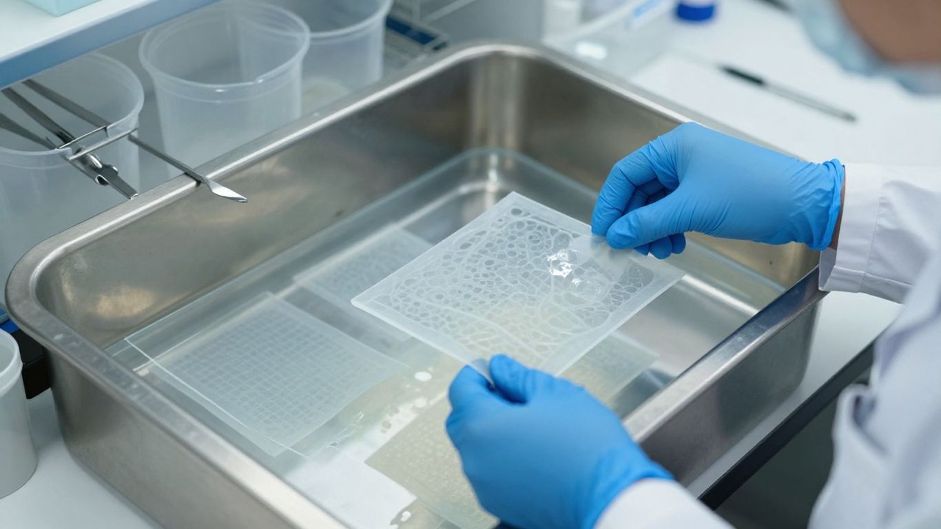
Mechanical delamination removes accessible layers such as serosa or mucosa when the anatomy allows them to be separated from the target matrix. The method should be consistent enough to avoid leaving thick cellular remnants, while gentle enough to prevent tearing or excessive compression. Operators need training, suitable instruments, and documented inspection points. Changes in force, angle, or tissue hydration can affect the final scaffold.
Freeze–thaw cycles for cell disruption
Freezing and thawing can disrupt cell membranes through ice formation and the repeated movement between temperature states. The number and temperature range of cycles should be established experimentally because excessive cycling may alter matrix structure, while insufficient cycling may leave intact cells. After treatment, the released debris must be removed rather than left within the scaffold. Physical methods such as freeze–thaw treatment are discussed further in this scaffold-production review.
Hypotonic or osmotic treatment considerations
Hypotonic exposure can promote water influx and cellular swelling, which may support membrane disruption. Duration, solution composition, temperature, and tissue thickness all influence the result. Overexposure can increase swelling or alter matrix properties, so you should monitor dimensions and appearance alongside cellular clearance. Osmotic treatment is best used as a controlled process variable, not as a universal shortcut.
Repeated rinsing and removal of cellular debris
Rinsing is the quiet work that determines whether disrupted material actually leaves the scaffold. Flow, agitation, volume, temperature, and cycle count should be standardized, with sampling used to confirm progress. A compact process record can help operators track the variables most likely to affect batch consistency:
- Tissue dimensions and starting mass
- Number and conditions of freeze–thaw cycles
- Osmotic exposure time and solution parameters
- Rinse volume, duration, and agitation
- Visual and analytical release observations
These records make deviations easier to investigate and help connect processing conditions with histological, biochemical, and mechanical results. Clear documentation is especially valuable when a scaffold appears clean macroscopically but retains microscopic debris.
Sterilization and packaging after processing
Sterilization must be selected with the final matrix in mind. The method should achieve the required microbial reduction without causing unacceptable changes in collagen, porosity, hydration, or biological activity. Packaging should protect the sterile barrier and preserve the product through storage and transport. You should validate the complete system, including sterilization, packaging integrity, shelf life, and handling instructions.
How to evaluate decellularization quality
No single test proves that a tissue is successfully decellularized. You need complementary evidence showing that cellular and nuclear material has been reduced, the ECM remains suitable, and the finished scaffold is sterile and biocompatible. Evaluation should compare processed tissue with an appropriate starting reference and include predefined acceptance criteria. The broad principles described in this ECM process overview are useful, but your final panel must match the product and intended use.
Histological evidence of cellular and nuclear removal
Histology can show whether nuclei, intact cells, and cellular remnants remain within the matrix. Use stains and imaging methods appropriate to the tissue, and examine multiple regions rather than relying on a single field. Pay attention to difficult-to-process interfaces, folds, edges, and thicker areas. A visually pale section is not enough unless the relevant cellular structures have been specifically assessed.
DNA and residual nucleic acid testing
Quantifying residual DNA or other nucleic acid material adds a biochemical measure to histological observations. Sampling plans should account for tissue heterogeneity and extraction efficiency. Results are most useful when interpreted alongside fragment size, distribution, and microscopic findings rather than treated as an isolated pass-or-fail number. The assay, controls, and reporting units should be fixed before routine release testing.
Extracellular matrix composition and protein profiling
Protein profiling helps determine whether processing preserved the matrix components that give the scaffold its biological identity. Depending on the application, you may examine collagen types, elastin, glycosaminoglycans, adhesion proteins, and selected growth-factor signals. Comparing composition before and after processing can reveal losses that are invisible in routine histology. The question is not only whether the tissue is acellular, but whether it still has the intended ECM composition.
Structural, mechanical, and morphological assessment
Microscopy, imaging, thickness measurements, swelling studies, and mechanical tests can reveal changes in architecture and handling behavior. You should assess whether the scaffold remains flexible, porous, cohesive, and appropriate for its intended site. A wound dressing and a load-bearing implant will not require the same mechanical profile. Test conditions should therefore reflect storage state, hydration, sterilization, and the way the material will be used.
Sterility, biocompatibility, and immunogenicity testing
Sterility testing and bioburden controls address microbial safety, while biocompatibility studies examine how the material interacts with cells and tissues. Depending on the intended use, evaluation may include cytotoxicity, sensitization, irritation, implantation response, and immunological endpoints. Animal or clinical evidence should be interpreted in the context of the final sterilized product, not an intermediate laboratory sample. This is where process quality and biological performance meet.
Benefits and limitations of the approach
An enzyme free strategy can be attractive when you want to preserve a tissue’s native organization and reduce the number of processing reagents. Its benefits are closely tied to the starting material and the quality of validation. It is not inherently superior for every tissue, application, or manufacturing scale. You should judge it against measurable outcomes: residual cellular content, matrix composition, mechanical behavior, sterility, and biological response.
Potential effects on inflammation and macrophage response
Residual cells, nucleic acids, damaged proteins, and contaminants can all influence host inflammation. Conversely, a well-preserved and adequately processed ECM may provide a more familiar biological environment for remodeling. Macrophage response is shaped by the scaffold, wound condition, sterilization, implantation site, and many other variables. Claims about a favorable response therefore require evidence from the specific material and model being evaluated.
Support for cell attachment, migration, and angiogenesis
Retained adhesion proteins and matrix architecture may give cells physical sites and biochemical cues for attachment and movement. A porous, hydrated structure can also support infiltration when its dimensions fit the target tissue. Angiogenesis is a complex biological outcome and should be demonstrated through appropriate in vitro or in vivo assays rather than inferred from composition alone. Scaffold design and wound-bed preparation remain equally relevant.
Processing trade-offs compared with detergent-based methods
The main trade-off is between simplicity and clearance power. Physical processing may reduce reagent exposure and preserve selected matrix features, but it can be slower, operator-sensitive, or less effective in dense tissues. Detergent-based processing can improve access to cellular material but may alter proteins, lipids, ultrastructure, or downstream cell behavior if not carefully controlled. A tissue-specific comparison is more meaningful than a blanket preference for one category.
Risks related to incomplete decellularization or contamination
The most serious risks include residual cellular material, microbial contamination, endotoxin, inconsistent thickness, and damage introduced during handling or sterilization. These risks can be reduced through source controls, validated parameters, environmental monitoring, and release testing. You should also define what happens when a batch fails: reprocessing may change the matrix, while disposal affects yield and cost. Quality planning belongs at the beginning of process development, not after a failure.
Applications and implementation considerations
Enzyme free decellularization is relevant wherever a natural ECM scaffold is expected to support repair, cell interaction, or tissue remodeling. Its clinical and research value depends on matching the scaffold to the wound or defect rather than treating “natural” as a complete specification. Shape, thickness, porosity, hydration, degradation, and handling all matter. The final application should guide both process development and evaluation.
Wound healing and soft-tissue scaffolds
A thin, flexible ECM can be useful when you need close contact with a wound bed and a surface that supports tissue repair. Potential use areas include soft-tissue defects, burns, chronic wounds, surgical wounds, and preclinical wound models, provided the material has evidence appropriate to the indication. CholeDerm is documented as a collagen-rich, sterile ECM scaffold derived from porcine cholecyst and intended for wound-care applications; its documented properties should not be generalized to unrelated matrices.
Use in preclinical tissue-regeneration studies
In preclinical work, you can use an enzyme free scaffold to study cell attachment, inflammatory signaling, vascular response, granulation, epithelialization, and remodeling. Experimental design should include appropriate controls, standardized wound dimensions, blinded assessment where possible, and prespecified endpoints. Report the processing method and sterilization conditions in enough detail for another laboratory to understand what was actually tested. A promising result in one animal model does not establish clinical effectiveness.
Matching scaffold properties to wound requirements
A scaffold for a shallow, exuding wound may need different absorbency and handling characteristics from one intended for a deeper soft-tissue defect. Consider wound depth, surface area, exudate, infection status, vascularity, mechanical movement, and the need for graft support. You should also plan how the material will be secured, covered, monitored, and replaced. The scaffold is one part of a broader wound-care pathway, not a replacement for assessment, debridement, infection control, or appropriate dressing selection.
Regulatory, manufacturing, and quality-system expectations
Moving from research material to a medical product requires control of raw materials, suppliers, processing, personnel, environment, equipment, packaging, sterilization, and records. Define critical process parameters and critical quality attributes early, then validate analytical methods and acceptance criteria. Manufacturing under an appropriate quality system supports traceability and change control. Clinical or preclinical evidence must correspond to the final product, its intended use, and its labeled conditions.
Conclusion
Enzyme free tissue decellularization offers a useful route for producing ECM scaffolds when tissue architecture and biological composition are worth protecting, but its success depends on tissue selection, controlled physical processing, thorough rinsing, validated sterilization, and multidimensional testing. If you match the workflow to the source tissue and the intended wound or regenerative application, you can make a more defensible decision about whether the approach provides the right balance of cellular clearance and matrix preservation.
Frequently Asked Questions
What is enzyme free tissue decellularization?
It is a decellularization approach that removes cellular material through physical, osmotic, and washing steps without adding enzymes such as proteases or nucleases. The aim is to retain the extracellular matrix while reducing potentially immunogenic cellular remnants.
Does enzyme free processing mean no chemicals are used?
No. Buffers, osmotic solutions, water, and sterilization agents may still be part of the workflow. The phrase specifically refers to avoiding added enzymes, and sometimes detergents as well, depending on the defined process.
Can every tissue be decellularized without enzymes?
No. Tissue thickness, cellularity, density, geometry, and matrix composition all affect feasibility. Some tissues may require additional processing or a different strategy to achieve adequate cellular removal.
Why is the extracellular matrix preserved?
The ECM provides structural support and may retain collagen, elastin, glycosaminoglycans, adhesion proteins, and matrix-associated signals. These features can influence cell attachment, migration, hydration, remodeling, and mechanical behavior.
How do you know whether a scaffold is adequately decellularized?
Use several tests together, including histology, nuclear staining, residual DNA analysis, protein or ECM profiling, structural and mechanical testing, sterility testing, and biocompatibility assessment. No single assay is sufficient for every application.
What are the main risks of an incomplete process?
Incomplete processing can leave cells, nuclei, DNA, or contaminants in the scaffold. These residues may increase inflammatory or immunological responses and can also create batch-to-batch variability or microbial safety concerns.
Is a preserved ECM automatically suitable for wound healing?
No. Suitability also depends on sterilization, biocompatibility, mechanical and handling properties, degradation, contamination control, clinical evidence, and the specific wound indication. A preserved matrix still needs application-specific validation.