
Key Takeaways
Your extracellular matrix does far more than fill space around cells. It provides structure, stores biological signals, and helps coordinate the movement, activity, and maturation of cells during repair.
- ECM supports overlapping phases of hemostasis, inflammation, proliferation, and remodeling.
- Collagen, elastin, glycosaminoglycans, proteoglycans, and adhesion proteins guide different aspects of cell behavior.
- Matrix architecture influences how fibroblasts, endothelial cells, and keratinocytes attach and migrate.
- Preserving bioactive ECM molecules may help maintain signals that support organized tissue repair.
- Research on porcine cholecyst-derived ECM includes wound, burn, diabetic, and soft-tissue models, but preclinical findings require careful interpretation.
What the extracellular matrix does during tissue repair
When tissue is injured, the extracellular matrix is damaged along with cells and blood vessels. Repair therefore requires more than closing the surface: your body must rebuild a temporary environment in which cells can attach, communicate, move, and produce new tissue. The ECM changes continuously as healing progresses, so its composition and physical properties matter at each stage.
The basic idea behind ECM and wound healing research is that the matrix acts as both a physical framework and a biological participant. It can be broken down, rebuilt, and remodeled in response to signals from platelets, immune cells, fibroblasts, endothelial cells, and epithelial cells.
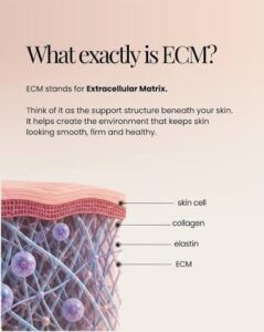
More than structural support
Collagen provides tensile strength, but it is only one part of the matrix. Elastin contributes recoil, glycosaminoglycans help retain water, and adhesion proteins give cells surfaces to grip. Together, these components create a three-dimensional setting that influences how cells spread and organize.
The matrix also helps anchor cells to surrounding tissue. That anchoring is not passive. The way a cell attaches can affect its shape, movement, survival, and production of new matrix. For you as a clinician or researcher, this is why a biological scaffold should not be judged only by whether it covers a wound.
How ECM creates a local healing microenvironment
A wound microenvironment includes oxygen availability, fluid balance, mechanical forces, inflammatory mediators, and matrix-derived signals. ECM components can bind or present signaling molecules close to the cells that need them. This local arrangement helps coordinate events that would be less orderly if the signals were dispersed without a supporting framework.
Matrix stiffness and architecture also influence cell behavior. A compliant, hydrated environment may support migration, while a denser structure can provide stronger resistance and attachment. These effects interact with growth factors and immune signals rather than operating in isolation.
The relationship between ECM breakdown and repair
Early matrix breakdown is necessary. Enzymes remove damaged material and open space for incoming cells, while fragments of matrix can influence inflammation and migration. The problem arises when degradation remains excessive or rebuilding does not keep pace, as can happen in chronic wounds.
Healthy repair depends on a changing balance between removal and replacement. Fibroblasts deposit new matrix, endothelial cells help establish blood supply, and epithelial cells move across the wound surface. If the matrix remains disorganized, cells may receive conflicting mechanical and biochemical cues.
Why tissue source and composition matter
A scaffold’s tissue of origin affects its collagen arrangement, accessory proteins, hydration, thickness, and potential interaction with host cells. Two materials described broadly as ECM may therefore behave differently because their native architecture and molecular profiles are not identical.
This is also why processing deserves attention. Decellularization and stabilization can improve handling or reduce cellular residues, but aggressive processing may alter the architecture and remove biologically active components. The relevant question is not simply whether a scaffold is natural; it is what structure and signals remain available after processing.
How ECM coordinates the stages of wound healing
Wound healing is often described in four phases: hemostasis, inflammation, proliferation, and remodeling. In practice, these stages overlap, and the ECM changes throughout the sequence. It first helps contain injury, then supports immune-cell activity, cell growth, vascular development, epithelial coverage, and finally the reorganization of repaired tissue.
The same matrix can therefore have different roles at different times. A provisional clot is useful early, while a more organized collagen network becomes important later. Understanding this progression is central to understanding the ECM in tissue repair, especially when you evaluate biological scaffolds for difficult wounds.
The provisional matrix during hemostasis
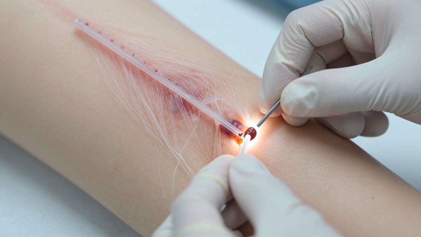
Immediately after injury, clotting creates a provisional matrix made largely from fibrin and associated blood components. It helps limit bleeding and provides an early surface for inflammatory cells and repair cells to attach. Platelets within the clot also release mediators that recruit cells and influence later matrix production.
This temporary structure is not meant to remain unchanged. As the wound moves toward proliferation, fibrin is replaced or supplemented by newly deposited matrix. Its early value lies in giving the wound a provisional organization when native tissue architecture has been disrupted.
ECM regulation of inflammation
Inflammatory cells enter the wound to remove debris, control contamination, and prepare the site for repair. ECM fragments and matrix-bound signals can influence how these cells behave, while immune cells in turn release enzymes and mediators that reshape the matrix.
A controlled inflammatory response clears the wound without keeping it locked in an inflammatory state. In contrast, persistent inflammation can damage newly deposited matrix, impair cell migration, and contribute to a chronic wound environment. The matrix is one of the places where these competing signals meet.
Supporting proliferation, granulation, and re-epithelialization
During proliferation, fibroblasts produce new matrix, endothelial cells contribute to vascular development, and keratinocytes move across the wound surface. Granulation tissue fills the defect and provides a transitional bed for continued repair. The ECM supports each process by offering attachment sites and by organizing local biochemical cues.
You can think of this phase as a coordinated construction process rather than simple tissue replacement. Cells need a surface, nutrients, signals, and room to move. A scaffold that retains useful architecture may support several of these requirements at once, although its effect still depends on the wound environment and clinical management.
Remodeling tissue into a stronger repair
Remodeling begins while proliferation is still underway and may continue for a prolonged period. Collagen is reorganized, cross-linked, and replaced according to mechanical demand. The repaired tissue gradually becomes more stable, though it may not fully reproduce the original tissue.
The quality of remodeling depends on the balance between matrix synthesis and degradation. Excessive or poorly organized collagen can contribute to fibrosis, while insufficient deposition can leave tissue weak. Organized remodeling matters because closure alone does not describe the final quality of repair.
How ECM components guide cell behavior
Cells do not respond to the ECM as if it were a single substance. They detect its fibers, binding sites, hydration, stiffness, and associated molecules through receptors and mechanical sensing systems. The combined signals help determine whether a cell remains attached, migrates, divides, changes phenotype, or produces more matrix.
For practical evaluation, it helps to separate the main matrix components while remembering that they work together. A collagen-rich scaffold without suitable adhesion molecules may behave differently from one that preserves a broader molecular profile.
Collagen as the structural framework
Collagen forms much of the tensile framework of connective tissue. Its fibers give cells a physical surface and help the developing repair tolerate mechanical forces. Different collagen types can contribute to organization, anchoring, and interaction with other matrix molecules.
As fibroblasts enter a wound, they both use and modify this framework. They deposit new collagen, contract the matrix, and respond to the density and alignment of existing fibers. The resulting structure influences how later remodeling proceeds.
Elastin and glycosaminoglycans in flexibility and hydration
Elastin helps tissues stretch and return toward their original shape. That property is especially relevant in skin and other tissues exposed to repeated movement. Glycosaminoglycans, meanwhile, bind water and contribute to a hydrated environment through which molecules and cells can move.
Hydration affects more than moisture alone. It changes diffusion, matrix swelling, and the mechanical feel of the scaffold. A balance between flexibility and structural stability can help a material conform to a wound without becoming biologically inert.
Proteoglycans and adhesion proteins in cell organization
Proteoglycans such as decorin, lumican, and mimecan can influence collagen organization and the presentation of signaling molecules. Adhesion proteins such as fibronectin, vitronectin, and periostin help cells attach and migrate across the matrix.
These molecules are easy to overlook when attention centers on collagen. Yet a cell needs both a framework and instructions for interacting with it. Their distribution can affect where cells settle and how efficiently they move through a scaffold.
Growth factors and matrix-bound signaling molecules
Growth factors including VEGF and FGF activity are associated with vascular development, granulation, and tissue repair. In native tissue, signals may be held near the matrix and released as the matrix is remodeled. This can provide spatial and temporal context that a freely dissolved signal may not have.
The presence of a molecule does not automatically establish a clinical effect. Its activity depends on preservation, concentration, release, receptor availability, and the condition of the wound. For that reason, protein profiling and functional studies are best considered together.
How ECM guides cells into damaged tissue
Cell recruitment is one of the clearest ways a scaffold can influence repair. Fibroblasts need to attach and move into the wound, endothelial cells need to participate in vascular development, and keratinocytes need to spread across the surface. Each cell type responds to different combinations of matrix structure and biochemical cues.
Architecture determines whether these cells can physically enter the material. A dense, poorly connected structure may limit infiltration, while a porous but unstable structure may fail to maintain a useful repair bed. The most relevant design is therefore tissue- and application-dependent.
Fibroblast attachment and migration
Fibroblasts attach to matrix proteins through adhesion receptors and then move along available fibers or surfaces. Once established in the wound, they produce collagen and other matrix components, helping replace the provisional environment with a more durable one.
Their behavior is affected by fiber alignment, hydration, stiffness, and the presence of adhesion proteins. A scaffold that supports attachment but restricts movement may not provide the same biological setting as one with interconnected pathways.
Endothelial cell recruitment and angiogenesis
Endothelial cells form the lining of new blood vessels and respond to local signals from hypoxia, inflammatory cells, growth factors, and the ECM. Their migration into a wound supports the delivery of oxygen and nutrients needed by metabolically active repair tissue.
Angiogenesis is not an isolated event. New vessels must integrate with granulation tissue and later remodeling. Matrix composition can help provide both a route for movement and a context for vascular signaling.
Keratinocyte movement during re-epithelialization
Keratinocytes at the wound edge loosen their attachments, migrate across the provisional bed, and gradually restore an epithelial barrier. They require a surface that supports movement while allowing them to maintain appropriate cell-cell organization as closure develops.
If the wound bed is excessively dry, inflamed, contaminated, or mechanically unstable, re-epithelialization may be delayed. ECM-based approaches are therefore only one part of care; wound preparation and management of the surrounding environment remain essential.
How scaffold architecture affects cellular infiltration
A scaffold’s thickness, porosity, flexibility, and fiber arrangement influence how cells enter and distribute through it. A thin, flexible, porous mesh may conform closely to an irregular wound and offer accessible paths for infiltration, while a thicker material may behave differently.
When you assess architecture, consider what the target cells must do rather than relying on a single material descriptor. The useful questions include whether cells can attach, whether they can move through the matrix, and whether the scaffold remains sufficiently stable while new tissue forms.
How ECM influences inflammation and tissue remodeling
Repair is shaped by the dialogue between matrix and immune cells. Macrophages, neutrophils, fibroblasts, and endothelial cells encounter matrix proteins and fragments while responding to injury. Their activity can help restore tissue or sustain damage, depending on timing and local conditions.
A biological scaffold should therefore be considered in terms of host-material interaction, not merely physical coverage. The goal is not to eliminate inflammation, which is necessary for defense and cleanup, but to support a transition toward repair.
Balancing inflammatory and reparative macrophage responses
Macrophages can adopt different functional states during healing, although the biology is more continuous than a simple two-category label. Early inflammatory activity helps clear debris and coordinate defense. Later reparative activity supports vascularization, matrix deposition, and remodeling.
Research on porcine cholecyst-derived ECM has evaluated macrophage phenotypes and reported evidence of an M2-dominant, low-inflammation response in the studied models. That finding should be read as model-specific evidence of host-material interaction, not as a guarantee that every wound will respond identically.
Why controlled inflammation supports healing
Inflammation becomes useful when it is proportionate and resolves as repair advances. It removes damaged material, recruits necessary cells, and prepares the matrix for new tissue. If it persists, protease activity and inflammatory mediators can damage the repair environment.
Clinical assessment remains central because infection, ischemia, diabetes, pressure, and repeated trauma can all alter the inflammatory course. A scaffold cannot compensate for every barrier to healing, and its role should be considered alongside wound-bed preparation and treatment of underlying conditions.
Reducing excessive fibrosis and disorganized repair
Fibrosis develops when repair signals favor persistent matrix deposition and contraction rather than restoration of balanced tissue structure. Excessive collagen, poor fiber organization, and prolonged inflammation can all contribute to a stiff or functionally limited scar.
Matrix composition may influence this outcome by affecting macrophage behavior, fibroblast activity, and the way collagen is deposited. Still, evidence must be interpreted carefully: a favorable remodeling signal in an animal or laboratory model does not establish the same result in every clinical setting.
The importance of preserving bioactive matrix signals
Processing can remove cellular material while also affecting proteins, glycosaminoglycans, growth-factor activity, and native architecture. Preserving a broader set of signals may help maintain the biological complexity that guides host cells.
This is one reason matrix characterization matters. Functional testing, protein profiling, degradation studies, and immune-response evaluation together provide a more useful picture than a simple statement that a material is collagen-based.
How biological scaffolds preserve and deliver ECM signals
A biological scaffold begins as tissue, but it becomes a medical material through preparation, decellularization, shaping, stabilization, and quality control. Each step can alter its mechanical properties and biological content. The central design challenge is to reduce unwanted cellular material while retaining enough architecture and signaling capacity to support repair.
That balance is not identical for every tissue source or application. A scaffold for a superficial wound may need flexibility and conformability, while a load-bearing application may require a different stability profile.
Why native architecture can influence scaffold performance
Native architecture creates the spatial arrangement of fibers, pores, and binding sites that cells encounter. Preserving that arrangement may support attachment and infiltration in ways that are difficult to reproduce with isolated proteins alone.
In porcine cholecyst-derived ECM research, the scaffold has been described as collagen-rich with a three-dimensional fibrous or mesh-like architecture. Such findings help explain why tissue origin and structural preservation are discussed together when evaluating a biological material.
Effects of decellularization on ECM composition
Decellularization aims to remove cells and cellular residues that could provoke an unfavorable host response. However, chemical, enzymatic, and mechanical treatments may also disrupt fibers or reduce biologically active molecules if they are too aggressive.
The outcome depends on the tissue and the method. You should therefore ask which residues were measured, which matrix components were retained, and whether the processed scaffold still supports the intended cellular response.
Non-detergent and non-enzymatic processing approaches
The PrisTINE™ process is described in the supplied technical material as a non-enzymatic, non-detergent approach intended to retain native ECM architecture and biologically active biomolecules. That description supports a discussion of processing strategy, not a claim that the resulting material is free of every chemical treatment or stabilization step.
Research on cholecyst-derived ECM also includes stabilization and crosslinking studies. These approaches can be used to examine degradation and handling, but their effects on biomolecule retention and host interaction must be evaluated for each formulation.
Balancing biocompatibility, degradation, and biomolecule retention
A scaffold must remain present long enough to support repair, yet it should also permit host tissue to replace or remodel it. Excessive persistence can interfere with integration, while rapid degradation may remove structural support too early.
The desired balance depends on wound depth, tissue mechanics, vascularity, and the intended role of the material. This is why degradation studies, cytocompatibility testing, immune-response analysis, and tissue integration findings need to be considered together rather than treated as interchangeable measures.
What research shows about cholecyst-derived ECM
Cholecyst-derived ECM has been investigated as a naturally derived scaffold because the gallbladder provides a relatively thin, collagen-rich tissue with a distinctive fibrous architecture. The research program includes material characterization, cell interaction, immune response, degradation, wound healing, and engineered formats.
The evidence is primarily preclinical. It can clarify mechanisms and identify promising patterns, but it does not by itself establish human efficacy, universal clinical indications, or identical performance across formulations.
The mesh-like structure of porcine cholecyst ECM
Studies describe porcine cholecyst ECM as having a tight, mesh-like collagen network with a three-dimensional fibrous architecture. Its thin and flexible character has been investigated in relation to wound conformity and cellular interaction.
These physical features may help explain why cells can attach to and move through the material in laboratory and animal studies. They should still be understood as material properties studied in specific preparations, not as a promise that every scaffold behaves the same way in every wound.
Reported proteins and signaling molecules in the scaffold
Research has evaluated collagen, elastin, sulfated glycosaminoglycans, VEGF, and bFGF activity in cholecyst-derived scaffolds. Protein-profiling work also identified ECM-associated proteins including fibronectin, nidogen, decorin, and lumican.
Taken together, these findings support viewing the scaffold as a biologically complex matrix rather than as collagen alone. The biological relevance of each component depends on how it is preserved, presented, and made available during degradation and host remodeling.
Findings from wound, burn, diabetic, and soft-tissue models
The supplied research includes rabbit full-thickness wound studies, a rabbit burn model, a diabetic-rat wound model involving a gelatin-modified cholecyst-derived scaffold, dog full-thickness lacerated wounds, and rat soft-tissue or muscle-defect models. Reported observations include granulation, epithelialization, angiogenic responses, scaffold integration, and organized remodeling in the evaluated models.
A compact view of the evidence helps keep each finding in context:
| Study context | Reported focus | Evidence status |
|---|---|---|
| Rabbit full-thickness wound | Granulation and epithelialization | Preclinical animal study |
| Rabbit burn wound | Inflammation and healing response | Preclinical animal study |
| Diabetic rat wound with gelatin-modified scaffold | Angiogenic response and closure | Specific experimental formulation |
| Dog full-thickness lacerated wound | Healing parameters and tissue repair | Naturally occurring animal wound model |
These results suggest potential across several repair environments, but they should not be converted into broad clinical guarantees. Model, formulation, wound type, and study design all affect interpretation.
Interpreting preclinical evidence and its clinical relevance
Preclinical research can show whether a scaffold supports cell attachment, vascular development, tissue infiltration, immune modulation, or remodeling under controlled conditions. It can also reveal safety signals and guide the design of later studies. It cannot replace well-designed clinical evidence in human patients.
For clinicians, the most useful approach is to connect the material’s documented properties with the wound’s needs while maintaining appropriate caution. The broader field’s ECM scaffold discussion in wound care can help frame why architecture and cell infiltration matter, but product selection still requires professional judgment and current product information.
Conclusion
How ECM Guides Tissue Repair is best understood as a story of structure, signals, cells, and timing: the matrix helps organize the wound environment while cells rebuild and remodel it. Cholecyst-derived ECM research adds a useful example of how tissue source, mesh-like architecture, molecular composition, and processing can shape scaffold behavior, while its preclinical findings should be interpreted with scientific care.
Frequently Asked Questions
What is the extracellular matrix?
The extracellular matrix is the network of proteins, polysaccharides, and associated molecules surrounding cells. It provides structural support while also influencing cell attachment, migration, signaling, and tissue organization.
How does ECM support wound healing?
ECM supports wound healing by providing a temporary or developing framework for cells, binding biological signals, and helping coordinate inflammation, proliferation, vascular development, epithelial coverage, and remodeling.
Why is collagen not the only important ECM component?
Collagen provides much of the structural framework, but elastin, glycosaminoglycans, proteoglycans, adhesion proteins, and growth-factor-associated signals also influence hydration, flexibility, cell attachment, migration, and organization.
What happens to ECM during inflammation?
The matrix is partly degraded and remodeled during inflammation. Its fragments and associated signals can influence immune-cell activity, while inflammatory cells release enzymes and mediators that prepare the wound for later repair.
How do cells enter an ECM scaffold?
Cells enter through available pores, fiber pathways, and surface attachment sites. Infiltration depends on architecture, porosity, thickness, hydration, stiffness, cell type, and the biological condition of the wound.
Can ECM scaffolds prevent scar formation?
No scaffold can be assumed to prevent scarring in every situation. ECM materials may influence inflammation and remodeling, but scar quality depends on wound depth, infection, blood supply, mechanical forces, patient factors, and the overall treatment plan.
What does preclinical evidence tell you about an ECM material?
Preclinical evidence can show how a material behaves in laboratory or animal models, including its interaction with cells, immune response, degradation, vascularization, and tissue remodeling. It does not automatically predict the same outcome in human clinical use.