
Key Takeaways
Collagen is central to repair, but it works within a larger biological system. Understanding that system helps you choose wound-healing support more thoughtfully.
-
Collagen provides structure, supports fibroblasts, and contributes to granulation tissue.
-
Elastin, glycosaminoglycans, proteoglycans, adhesion proteins, and growth factors add flexibility, hydration, signaling, and organization.
-
The extracellular matrix influences how fibroblasts, endothelial cells, keratinocytes, and macrophages behave.
-
Chronic inflammation, poor angiogenesis, excess proteases, infection, pressure, and exudate can undermine collagen-based support.
-
A biologically active, three-dimensional scaffold may offer structural cues that isolated collagen cannot provide on its own.
Collagen’s essential role in tissue repair
Collagen is one of the main structural proteins in skin and a major component of the extracellular matrix. When tissue is injured, it helps create a framework through which cells can move, attach, and rebuild the wound. That makes collagen highly relevant to wound care, but it does not make collagen the entire healing environment. To understand why collagen alone may not be enough, you first need to see what it does well.
How collagen supports the wound-healing scaffold
After injury, the wound develops a temporary matrix that supports incoming cells. Collagen later becomes part of the more durable framework, giving granulation tissue structure and helping the wound develop mechanical strength. Its fibers also interact with cell receptors and surrounding matrix molecules, so collagen can provide both physical support and biological cues.
For a broader overview of collagen-based dressings and other medical uses, see this collagen wound-care overview. The useful question is not simply whether collagen is present, but whether the surrounding wound environment allows cells to use it effectively.
Collagen production during the proliferative phase
During proliferation, fibroblasts produce collagen and other matrix components as granulation tissue fills the wound. At the same time, endothelial cells form new capillaries and keratinocytes begin moving across the surface. These events are connected: a developing matrix must support cells while the wound is still changing.
You can think of collagen as a framework under construction rather than a finished repair. Its value depends on fibroblast activity, available oxygen and nutrients, appropriate moisture, and coordinated signaling from growth factors and cytokines.
Why collagen quality and organization matter during remodeling
Remodeling continues for weeks or months. Early collagen, including type III collagen, is reorganized and progressively replaced or reinforced by stronger type I collagen. Fibers become more organized, while myofibroblasts contract the wound and vascular density gradually declines.
The arrangement of collagen therefore matters as much as its presence. A disorganized or rapidly degraded matrix may provide limited support, whereas a stable but remodelable structure can give cells time to attach, migrate, and contribute to stronger tissue.
When adding collagen may support a slow-healing wound
Collagen dressings may be considered for slow-healing wounds, ulcers, and surgical wounds, particularly when the wound bed needs matrix support. Their intended role is commonly described as stimulating fibroblasts, supporting matrix formation, and promoting granulation. They are not a substitute for assessment of perfusion, infection, pressure, nutrition, or the cause of delayed healing.
A practical review of collagen’s structural and signaling roles is available in this collagen healing guide. For you and the clinical team, the decision should be tied to the wound’s current phase and condition rather than to collagen as a stand-alone solution.
The other components that make up a functional extracellular matrix
The extracellular matrix is a network, not a single ingredient. Alongside collagen, it contains elastic proteins, hydrated carbohydrate-rich molecules, adhesion proteins, and signaling molecules. Together, these components influence the physical properties of the wound bed and the behavior of cells entering it.
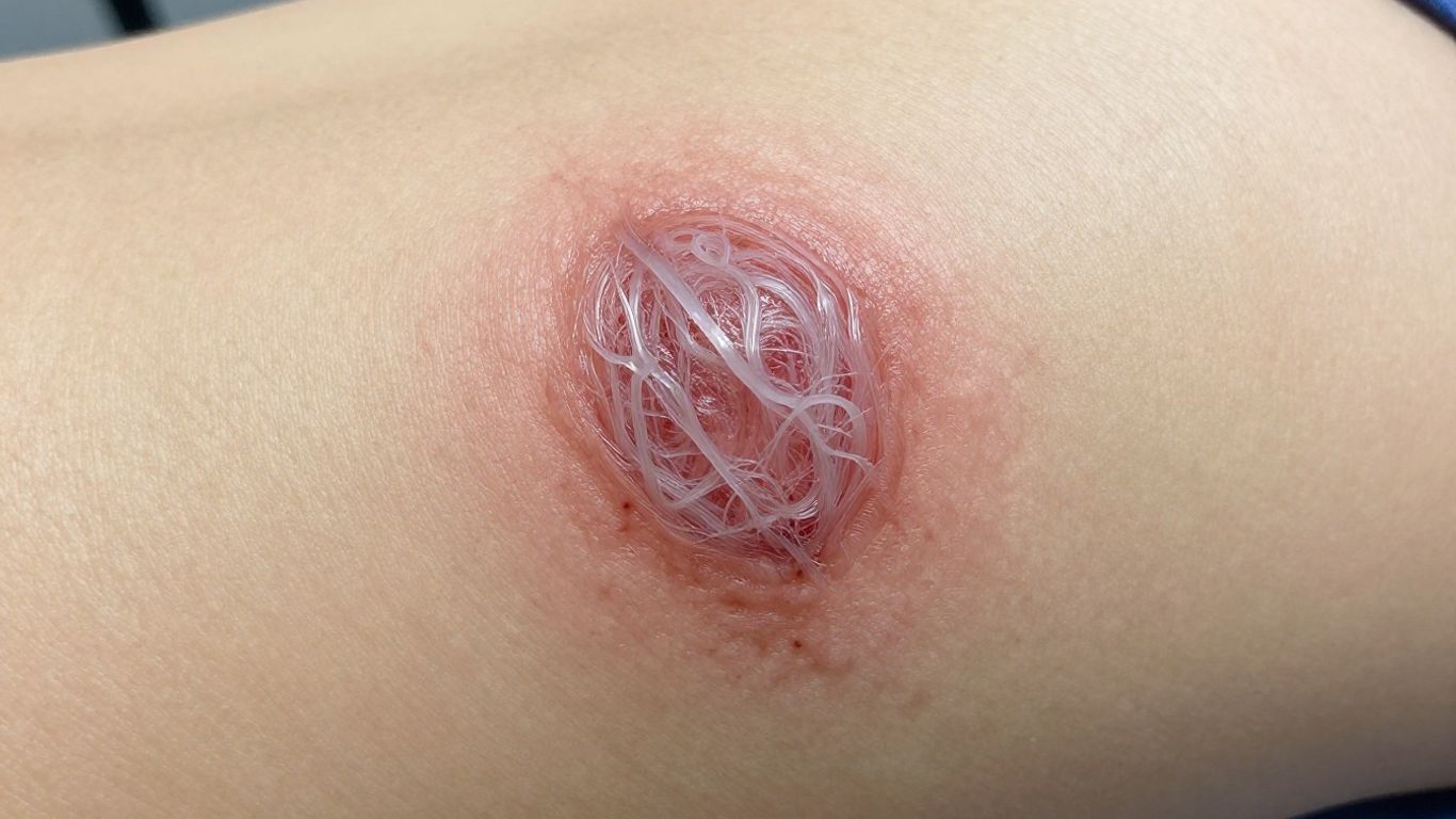
A useful way to picture the matrix is as both a building material and a local communication system. The following image captures that layered view of a healing scaffold.
Elastin and flexibility in repairing skin
Elastin helps tissues stretch and return toward their original shape. In repairing skin, that flexibility matters because a wound must tolerate movement while new tissue is still organizing. Collagen supplies much of the tensile framework, while elastin contributes resilience and helps the tissue respond to mechanical forces.
A matrix with both structural strength and flexibility is better suited to skin than a rigid material alone. The balance is especially relevant around joints, mobile skin, and wounds exposed to repeated tension.
Glycosaminoglycans and proteoglycans for hydration and signaling
Glycosaminoglycans attract and hold water, helping create a hydrated space around cells. Proteoglycans can bind growth factors and influence how those signals are presented, retained, or released. This gives the matrix a role in regulating the local chemical environment, not merely filling space.
Hydration must still be controlled. Excess fluid can macerate surrounding skin, while insufficient moisture can impair cell movement and epithelialization. The goal is a balanced wound bed rather than a uniformly wet or dry surface.
Fibronectin and adhesion proteins that guide cell movement
Fibronectin and related adhesion proteins help cells attach to the matrix and move across it. They provide binding sites that can guide fibroblasts, endothelial cells, and epithelial cells toward areas requiring repair. Without suitable adhesion cues, a wound may contain useful material but still fail to organize cell behavior effectively.
This is one reason matrix complexity matters. A scaffold containing several complementary adhesion and structural signals may interact with cells differently from a preparation consisting primarily of isolated collagen.
Growth factors and cytokines that coordinate repair
Growth factors and cytokines coordinate inflammation, proliferation, angiogenesis, matrix formation, and epithelialization. Examples include PDGF, FGF, EGF, TGF-beta, and IGF-1, each with overlapping but distinct roles. Their concentration, timing, and balance can be as important as their presence.
A chronic wound may show prolonged inflammation, reduced growth-factor activity, and elevated matrix metalloproteinases. That combination can prevent the matrix from becoming a stable platform for repair, even when collagen is supplied.
How the extracellular matrix directs healing cells
A wound-healing scaffold is biologically useful only if cells can interact with it. The matrix presents attachment sites, physical pathways, mechanical cues, and stored signals that affect how cells migrate and mature. Those interactions help determine whether repair progresses or stalls.
The same matrix can also influence different cell types at different stages. Fibroblasts build tissue, endothelial cells support vascularization, keratinocytes restore the surface, and macrophages help regulate inflammation and remodeling.
Supporting fibroblast attachment and migration
Fibroblasts enter the wound and attach to matrix proteins before producing collagen and other components. They need a surface that permits movement and a local environment that supports survival and activity. If the scaffold is too unstable, it may disappear before cells can organize it; if it is too dense, infiltration may be limited.
Fibroblast behavior is therefore shaped by both chemistry and architecture. A three-dimensional matrix can provide routes and attachment points that a thin layer of isolated protein may not reproduce.
Encouraging endothelial cells and new blood-vessel formation
Endothelial cells form new capillaries during proliferation, a process called angiogenesis. New vessels bring oxygen and nutrients and help remove waste, making vascularization essential for deeper or larger wounds. Matrix-associated signals such as VEGF and FGF can participate in this process.
Preclinical work on cholecyst-derived extracellular matrix has investigated endothelial-cell migration, tube formation, and angiogenic responses. These findings are useful for understanding mechanism, but animal and laboratory results should not be treated as proof of the same outcome in every patient.
Guiding keratinocyte movement and re-epithelialization
Keratinocytes migrate across the wound surface to restore the epidermal barrier. They need a wound bed that is moist enough for movement, protected from excessive trauma, and free from barriers such as necrotic tissue or uncontrolled bioburden. Matrix proteins can provide tracks and attachment cues during this process.
Re-epithelialization is not the same as complete healing. The deeper matrix, vascular network, inflammatory response, and remodeling phase still need to progress after the surface begins to close.
Influencing macrophage behavior and inflammation
Macrophages clear debris and coordinate the transition from inflammation to repair. Their behavior is influenced by signals from damaged tissue, microbes, cytokines, and the matrix itself. A prolonged inflammatory state can keep the wound from moving into productive proliferation.
For this reason, the immunological behavior of a scaffold deserves attention alongside its tensile or absorptive properties. The desired outcome is not to eliminate inflammation entirely, but to support a controlled response that permits regeneration and remodeling.
Why collagen alone may fall short in chronic wounds
Chronic wounds are not simply wounds with too little collagen. They often remain trapped in an inflammatory state, with impaired angiogenesis, poor cell migration, elevated protease activity, and recurrent microbial problems. Pressure, neuropathy, vascular disease, diabetes, and excess exudate can add further obstacles.
In that setting, adding a structural protein may address only one part of the problem. You must first understand what is preventing the wound from progressing through the normal phases of repair.
Prolonged inflammation and impaired cellular responses
Acute inflammation helps remove bacteria and damaged tissue. In a chronic wound, however, inflammation may persist and continue damaging the local matrix. Fibroblasts may respond poorly, macrophages may remain in a damaging inflammatory pattern, and the signals needed for proliferation may become unbalanced.
This creates a difficult feedback loop: damaged matrix limits cell activity, and reduced cell activity prevents the wound from rebuilding a healthier matrix. A scaffold may help, but only as part of a wider plan that addresses the cause of inflammation.
Poor angiogenesis and limited oxygen delivery
New tissue requires an adequate blood supply. Peripheral vascular disease, diabetes, edema, and pressure can reduce oxygen delivery or make the local environment unsuitable for capillary growth. Without sufficient perfusion, fibroblasts and keratinocytes may not function effectively even when a matrix is available.
Assessing circulation and relieving avoidable pressure can therefore be more urgent than selecting a collagen product. A biologically active dressing cannot compensate for every systemic or mechanical limitation.
Excess proteases and breakdown of the wound matrix
Matrix metalloproteinases and other proteases help remodel tissue, but excessive activity can break down newly deposited matrix before it becomes functional. Chronic wounds may therefore lose growth factors, adhesion proteins, and structural components faster than they can replace them.
The result is a wound bed with poor biological stability. Controlling the underlying inflammation and protecting the matrix may matter as much as supplying additional material.
Biofilm, infection, pressure, and uncontrolled exudate
A wound can also fail because of biofilm, infection, repeated pressure, shear, or poorly controlled exudate. These problems interfere with cell migration and may damage surrounding skin. Before you judge a scaffold or dressing, check whether the wound bed is prepared to receive it.
Several practical priorities often need to be addressed together:
-
Remove necrotic or nonviable tissue when clinically indicated.
-
Assess and manage infection or suspected biofilm.
-
Relieve pressure, friction, and repetitive mechanical stress.
-
Control exudate while protecting the surrounding skin.
These steps do not replace biological support; they create the conditions in which biological support has a reasonable chance to work. The dressing choice should follow that assessment, not stand in for it.
From collagen products to biologically active scaffolds
The central distinction is between isolated collagen and a tissue-derived extracellular matrix that preserves several structural and biochemical features. A three-dimensional scaffold may contain collagen alongside elastin, glycosaminoglycans, adhesion proteins, and matrix-associated signals. Its architecture can influence how cells attach, enter, and remodel the material.
This does not mean every complex scaffold is automatically better. Processing, source tissue, stability, biocompatibility, degradation, and clinical context all affect performance. The relevant question is whether the scaffold’s properties fit the wound environment.
How a three-dimensional matrix differs from isolated collagen
Isolated collagen can provide a useful structural substrate, but it may not retain the full arrangement of a native tissue matrix. A three-dimensional scaffold offers thickness, interconnected spaces, and multiple molecular cues that can support cell attachment and infiltration.
The distinction is especially important when you are considering how a material behaves over time. A scaffold is not merely a collagen dose; it is a temporary environment that may guide several cell populations during repair.
Why native architecture can affect cell infiltration and remodeling
Native architecture affects pore spaces, fiber arrangement, flexibility, and the distribution of matrix-associated molecules. These features can influence whether cells remain on the surface or migrate into the scaffold. They may also affect how the material is remodeled as new tissue develops.
Research on cholecyst-derived matrices has examined mesh-like architecture, cellular attachment, host-tissue infiltration, and in-vivo degradation. Such findings support the broader principle that structure and composition should be considered together.
Balancing scaffold stability with controlled degradation
A scaffold must remain present long enough to support repair, but it should also permit gradual remodeling. Excessive degradation can remove the framework too early, while excessive stability may interfere with tissue replacement or produce an undesirable foreign-body response.
Crosslinking and other stabilization approaches can alter degradation behavior, mechanical properties, and the retention of biomolecules. Those engineering decisions require careful validation rather than a simple assumption that more stability is always beneficial.
What research on cholecyst-derived ECM suggests about matrix complexity
CholeDerm® is described in the supplied research as a porcine cholecyst-derived acellular dermal matrix containing collagen, elastin, glycosaminoglycans, growth factors, and other matrix-associated proteins. Preclinical studies have investigated its use in rabbit, rat, and dog wound models, including full-thickness, burn, diabetic, and naturally occurring traumatic wounds.
The findings suggest that a preserved, biologically complex matrix can support cell interaction, angiogenic activity, granulation, and remodeling in the evaluated models. They remain preclinical findings, however, and should not be presented as a guarantee of clinical performance or as evidence that every experimental formulation is identical to a commercial product.
Choosing wound-healing support based on the wound environment
Choosing support begins with the wound, not with a favorite material. You need to consider exudate, tissue viability, depth, perfusion, infection risk, pressure, surrounding skin, and the patient’s broader health. A biologic scaffold may be appropriate in one setting and poorly timed in another.
The following comparison helps connect common wound-bed needs with broad dressing functions. It is a starting framework, not a substitute for clinical assessment.
|
Wound-bed need |
Supportive dressing characteristic |
Practical purpose |
|---|---|---|
|
Moderate to heavy exudate |
Gel-forming or absorbent material |
Manage fluid and reduce maceration risk |
|
Pressure or mechanical stress |
Cushioning and conformability |
Protect the wound and surrounding tissue |
|
Slow-healing wound with limited matrix support |
Collagen or biologic matrix |
Support fibroblasts, granulation, and tissue organization |
|
Irregular or cavity wound |
Moldable paste or powder |
Fill spaces while managing exudate |
|
Need for a biologically active interface |
ECM or other biologic dressing |
Provide structural and signaling cues |
The best choice may change as the wound progresses. Reassessing the wound bed is part of treatment, not an administrative step.
Matching dressings to exudate and wound-bed conditions
Hydrofiber dressings are used for moderate to heavy exudate and form a gel that conforms to the wound bed. Foam dressings can absorb excess exudate while maintaining moisture and providing cushioning. Collagen products are generally considered for slow-healing wounds, ulcers, and surgical wounds when matrix support is relevant.
You should also consider whether the material can be removed without disturbing granulation tissue and whether the surrounding skin is protected from moisture-associated damage. Exudate management and biological support are often complementary rather than competing goals.
Combining biological signals with moisture and exudate control
A biologic matrix may provide signals and a physical interface, while another dressing or therapy manages fluid. In the supplied research, CholeDerm® has been discussed in combination with negative-pressure wound therapy as a biological interface paired with mechanical exudate control and wound contraction.
The practical approach is to preserve contact between the matrix and wound bed while allowing fluid to leave safely. High-exudate wounds may require fenestration or other technique decisions made according to the applicable clinical protocol.
When debridement, infection management, or pressure relief comes first
A dressing cannot correct an untreated source of tissue damage. Nonviable tissue, infection, pressure, ischemia, edema, and repeated trauma may need attention before a biologic scaffold can contribute meaningfully. In some cases, cleansing, debridement, offloading, vascular assessment, or infection management is the immediate priority.
Your assessment should also include diabetes, neuropathy, immobility, medication history, and previous wound infections. Addressing these factors can change the wound environment more substantially than changing the dressing alone.
Interpreting preclinical ECM evidence without overstating clinical claims
Preclinical studies are valuable because they reveal how a material interacts with cells and tissue in controlled models. They can show evidence of angiogenesis, macrophage responses, granulation, epithelialization, or remodeling. They cannot, by themselves, establish effectiveness across human wound types or replace current product information and clinical judgment.
The research behind the CholeDerm® platform includes work on natural matrix preservation and cellular interaction, while the patented PrisTINE™ process is described as a non-enzymatic, non-detergent approach. When you read such evidence, keep the formulation, model, comparator, endpoint, and study limitations visible.
Conclusion
Collagen remains an essential part of tissue repair, but wound healing depends on a coordinated extracellular matrix, responsive cells, adequate perfusion, controlled inflammation, and a well-managed wound bed. When you evaluate collagen or an ECM scaffold, consider the whole environment: structure, signals, degradation, exudate, infection, pressure, and the evidence supporting the specific material and use.
Frequently Asked Questions
Is collagen important for wound healing?
Yes. Collagen provides structural support, contributes to granulation tissue, helps cells attach and migrate, and is reorganized during remodeling. Its effectiveness depends on the wider wound environment.
Why might collagen alone be insufficient in a chronic wound?
Chronic wounds may have prolonged inflammation, poor blood supply, excess protease activity, infection, biofilm, pressure, or uncontrolled exudate. Supplying collagen does not automatically correct these barriers.
What else is found in the extracellular matrix?
The matrix includes elastin, glycosaminoglycans, proteoglycans, fibronectin, other adhesion proteins, growth factors, and cytokines. These components influence flexibility, hydration, cell attachment, signaling, and remodeling.
How does the matrix affect healing cells?
It provides physical pathways and molecular cues that influence fibroblast attachment, endothelial-cell activity, keratinocyte migration, and macrophage behavior. Its architecture and composition both matter.
When might a collagen dressing be considered?
Collagen dressings may be considered for selected slow-healing wounds, ulcers, and surgical wounds when matrix support is clinically appropriate. The wound should still be assessed for perfusion, infection, pressure, exudate, and tissue viability.
What is the difference between isolated collagen and an ECM scaffold?
Isolated collagen supplies a structural protein, while an ECM scaffold may preserve a three-dimensional architecture and several matrix components. The clinical relevance depends on processing, biocompatibility, degradation, and the specific wound context.
Does preclinical ECM research prove that a scaffold will work in every patient?
No. Laboratory and animal studies can clarify mechanisms and show potential, but they do not guarantee outcomes in humans. Clinical decisions should rely on the available product information, evidence, and professional assessment.