
Key Takeaways
ECM is more than a structural layer: it creates a changing local environment that helps healing cells attach, move, communicate, and mature.
-
Collagen fibers provide structure, traction, and a path for tissue ingrowth.
-
Glycosaminoglycans, proteoglycans, adhesion proteins, and growth factors add biological signals.
-
Different cell populations read and respond to the matrix in different ways.
-
Controlled degradation allows the wound to replace the scaffold with organized tissue.
-
Cholecyst-derived ECM is being studied as a biologically complex scaffold in several preclinical models.
What the ECM microenvironment means in wound healing
When you ask how ECM provides a microenvironment for healing cells, the answer begins with its combination of physical structure and biological information. The matrix surrounds cells, gives them attachment points, and holds signals that can influence movement and activity. It also changes as the wound progresses, so the environment is not static.
From passive support to active biological signaling
You can think of ECM as the neighborhood in which repair takes place. Collagen and elastin help organize space, while glycosaminoglycans and proteoglycans bind water and signaling molecules. Together, these features influence whether a cell remains attached, migrates across the wound, divides, or begins producing new tissue.
That is why a scaffold cannot be judged only by its ability to cover a defect. Its chemistry, architecture, and rate of breakdown all shape the signals available to resident and incoming cells.
The difference between a scaffold and a microenvironment
A scaffold is often described as a physical framework. A microenvironment includes that framework, but also the local moisture, mechanical cues, adhesion molecules, growth factors, and immune signals surrounding the cells.
This broader view aligns with the ECM role in tissue repair, where the matrix is described as a three-dimensional setting that guides cell behavior while binding signaling molecules. In practice, the distinction helps you evaluate whether a material merely occupies space or actively supports coordinated repair.
How the wound changes the surrounding ECM
After injury, enzymes begin breaking down damaged matrix and provisional matrix components appear. Fibroblasts then deposit new collagen and other matrix molecules, while immune cells and endothelial cells alter the chemical environment around them.
In a healthy repair process, breakdown and rebuilding overlap. If degradation is excessive, the wound may lose structural cues faster than it can replace them. If rebuilding is disorganized, the result may be stiff scar tissue rather than mature, functional tissue.
Why the microenvironment matters for coordinated repair
Healing cells do not act independently. Fibroblasts need a suitable surface for matrix deposition, keratinocytes need a path across the wound, endothelial cells need cues for vascular ingrowth, and macrophages help determine whether inflammation persists or resolves.
A useful microenvironment lets these activities occur in sequence and in parallel. That coordination is one reason collagen alone is not the whole story; the broader ECM composition includes adhesion proteins, elastin, glycosaminoglycans, proteoglycans, and signaling molecules.
How ECM architecture guides cell behavior
The geometry of ECM affects what cells can reach and how they move through a wound. Fiber diameter, pore size, orientation, thickness, and stiffness all influence cell attachment and traction. These features also affect how fluid and signaling molecules move through the scaffold.
A biologically active matrix therefore has both a material side and a cellular side. Its architecture must be open enough for infiltration while remaining coherent enough to support the wound during early repair.
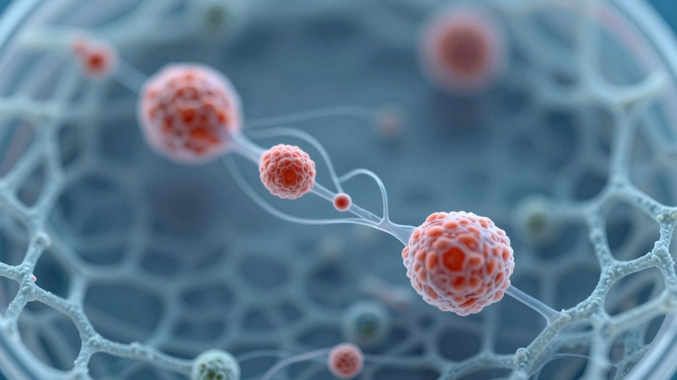
Collagen networks provide structure and traction
Collagen fibers form much of the tensile framework in connective tissue. When a fibroblast attaches to collagen, it can pull against the fibers, spread, and organize newly deposited matrix. This traction helps cells sense their surroundings and orient their movement.
The arrangement matters as much as the presence of collagen. A preserved network can provide more useful guidance than a fragmented collection of isolated collagen molecules.
Porosity and fiber organization support cell infiltration
Pores create routes through which cells, fluid, and soluble signals can move. If spaces are too small, infiltration may be limited; if the structure is too loose, cells may lack enough contact and support.
In a mesh-like scaffold, you can examine several practical questions:
-
Can cells enter the material rather than remain only on its surface?
-
Can wound fluid and signaling molecules move through the matrix?
-
Does the fiber arrangement provide multiple attachment paths?
-
Can the structure remain coherent while host tissue grows into it?
These questions connect architecture with function. Porosity is not useful in isolation; it has value when it supports the cellular activities required for repair.
Mechanical properties influence cell attachment and movement
Cells respond to stiffness, stretch, and resistance. A matrix that is too soft may not provide enough traction, while one that is excessively rigid can encourage abnormal tension and remodeling. Mechanical cues interact with biochemical signals, so the same molecule may produce different effects in different physical settings.
For you as a clinician or researcher, this means that handling characteristics and biological performance should not be considered unrelated. The way a scaffold conforms to a wound can influence how cells contact it and how evenly the wound bed is supported.
Degradation opens space for new tissue formation
Degradation is not simply a loss of material. Controlled breakdown can expose new attachment sites, release matrix-associated signals, and make room for host tissue. The timing is critical: degradation that occurs too quickly may remove support, while degradation that is too slow may restrict remodeling.
Crosslinking and other stabilization strategies are therefore studied as ways to adjust persistence. The goal is not permanent replacement, but a scaffold that remains present long enough to guide early repair and then yields to newly formed tissue.
How ECM biomolecules communicate with healing cells
The matrix contains several classes of molecules, each contributing a different kind of information. Adhesion proteins help cells attach, glycosaminoglycans organize water and signals, and growth factors influence proliferation and vascular responses. Proteoglycans add another layer by controlling how molecules are presented and retained.
This molecular diversity helps explain why a complex biological matrix may behave differently from a material made primarily from one purified component.
Adhesion proteins help cells attach to the scaffold
Fibronectin, vitronectin, and related proteins provide binding sites for cell-surface receptors. Once attached, a cell can spread, generate traction, and begin responding to nearby signals. Attachment also affects survival; cells generally behave differently when they can establish stable contact with an appropriate matrix.
The quality of those interactions depends on whether the proteins remain available in a useful arrangement. Processing that removes or alters them may change how cells recognize the scaffold.
Glycosaminoglycans regulate hydration and signaling
Glycosaminoglycans, or GAGs, are water-attracting molecules that contribute to the hydrated character of connective tissue. They can also bind growth factors and influence how those signals diffuse through the wound.
That dual role makes GAGs biologically important rather than merely decorative. The role of GAGs in regenerative medicine explains how they help turn ECM into an active signaling environment that supports granulation and angiogenesis.
Growth factors support proliferation and angiogenesis
Growth factors can encourage cells to divide, migrate, or adopt repair-related behaviors. In particular, vascular signals help endothelial cells form new vessels, which can improve the delivery of oxygen and nutrients to developing tissue.
Their effect depends on dose, timing, binding, and the condition of the wound. A matrix that retains or presents such signals may provide a more localized cue than a freely diffusing signal alone, although the biological outcome still depends on the wider wound environment.
Proteoglycans influence organized tissue remodeling
Proteoglycans combine a core protein with attached GAG chains. This structure allows them to interact with collagen, growth factors, and cell receptors. Decorin, lumican, and related molecules can influence fibril organization and the quality of matrix deposition.
That is one reason you should distinguish organized remodeling from simply producing more collagen. Tissue quality depends on how new fibers are arranged, crosslinked, and integrated with the surrounding matrix.
How ECM supports different cell populations
A wound contains many cell types, and each reads the matrix through its own receptors and signaling pathways. Fibroblasts build connective tissue, keratinocytes restore the surface, endothelial cells establish vascular supply, and macrophages help coordinate the inflammatory transition.
The matrix does not direct every cell in the same way. Instead, it offers overlapping physical and biochemical cues that are interpreted according to cell type and wound stage.
Fibroblasts and the formation of new connective tissue
Fibroblasts attach to matrix fibers, migrate into the wound, and produce collagen and other connective-tissue components. They also respond to stiffness and tension, which can influence whether repair remains organized or trends toward excessive contraction and fibrosis.
A supportive ECM gives fibroblasts places to attach and a structure around which new matrix can form. Their activity is most useful when deposition is followed by orderly remodeling rather than unchecked accumulation.
Keratinocytes and re-epithelialization
Keratinocytes must migrate across the wound surface before they can restore an epithelial barrier. They need a moist, permissive interface and appropriate adhesion cues, but they also need enough freedom to move rather than become tightly trapped.
This is why wound-bed preparation and moisture management remain essential even when a biological scaffold is used. The material supports the environment; it does not remove the need to address necrotic tissue, infection, perfusion, or exudate.
Endothelial cells and vascular ingrowth
Endothelial cells respond to vascular signals and migrate through suitable matrix spaces. Their activity can lead to new capillary formation, bringing oxygen and nutrients into developing granulation tissue.
Vascularization is especially relevant in wounds where perfusion is impaired. Still, a scaffold cannot compensate for every systemic or vascular problem, so you must interpret angiogenic findings alongside wound assessment and broader clinical care.
Macrophages and the transition from inflammation to repair
Macrophages are highly adaptable immune cells. Early inflammatory activity helps clear damaged material, while later reparative activity supports matrix deposition, vascular responses, and tissue maturation.
The matrix can influence this transition through its composition, degradation products, and physical presentation. The aim is not to eliminate inflammation, which is necessary at the beginning, but to support a timely shift toward repair.
How ECM helps regulate inflammation and remodeling
Inflammation and remodeling are not separate chapters of healing. They overlap, and the matrix participates in both. Damaged ECM can release signals, immune cells can alter matrix turnover, and newly deposited matrix can change the mechanical environment.
The quality of repair depends on maintaining enough inflammation to protect the wound while preventing a prolonged response that damages healthy tissue.
Balancing inflammatory and regenerative immune responses
A wound that remains dominated by inflammatory signals may show poor cell migration, impaired angiogenesis, and ongoing tissue breakdown. A more reparative immune environment can support granulation and matrix deposition, but it still needs regulation rather than unchecked activation.
You can therefore view immune modulation as a matter of timing and balance. The matrix is one influence among many, alongside infection control, perfusion, metabolic health, and mechanical stress.
Why controlled degradation matters for healing
As ECM breaks down, it creates space and may expose bioactive fragments. At the same time, excessive protease activity can destroy newly deposited matrix and signaling molecules. This imbalance is common in difficult-to-heal wounds.
A scaffold with a controlled degradation profile can provide temporary continuity while host cells repopulate it. That is a design objective, not a guarantee of a particular clinical outcome.
How ECM composition can influence fibrosis
Fibrosis is associated with excessive or poorly organized matrix deposition. Collagen quantity matters, but so do fiber alignment, crosslinking, proteoglycan interactions, mechanical tension, and the persistence of inflammatory signals.
Native elastin and other matrix components may contribute to a more familiar repair setting, although preservation alone cannot ensure normal tissue regeneration. The role of elastin in skin repair provides useful context for how elasticity and matrix organization affect healing.
The relationship between matrix turnover and tissue maturation
Early repair often produces provisional matrix that is later reorganized into stronger, more specialized tissue. Enzymes remove older components while fibroblasts adjust the composition and alignment of new fibers.
Maturation is gradual. A wound can appear closed while its matrix is still changing, which is why surface closure and restoration of tissue quality should not be treated as identical endpoints.
How cholecyst-derived ECM creates a healing environment
Porcine cholecyst-derived ECM has been investigated as a collagen-rich biological scaffold with a three-dimensional fibrous or mesh-like architecture. Research has examined its structure, biomolecule content, cell interactions, tissue response, and degradation. These findings make it a useful example of how tissue source can shape scaffold design.
The evidence remains primarily preclinical, so you should distinguish material characterization and animal-model findings from established human clinical outcomes.
The mesh-like architecture of porcine cholecyst ECM
Studies of cholecyst-derived ECM describe a fibrous, mesh-like structure that can provide attachment surfaces and pathways for infiltration. Its physical form is relevant because cells interact with an arrangement of fibers, not only with isolated collagen molecules.
The architecture has been investigated in relation to cellular attachment, proliferation, host-tissue infiltration, and remodeling. Those are mechanisms of interest, not proof that every wound will respond in the same way.
Collagen, elastin and glycosaminoglycans in the matrix
Characterization studies evaluated collagen, elastin, sulfated GAGs, VEGF, and basic fibroblast growth factor activity in cholecyst-derived material. Protein profiling also identified ECM-associated proteins such as fibronectin, nidogen, decorin, and lumican.
Taken together, these findings support describing the scaffold as biologically complex rather than as collagen alone. That distinction matters when you consider hydration, adhesion, signaling, and remodeling as connected functions.
Preserving native biomolecules during ECM recovery
The research includes a non-detergent and non-enzymatic approach for recovering cholecyst-derived ECM, alongside separate work on stabilization and crosslinking. The processing objective is to remove cellular material while retaining as much native architecture and biologically relevant matrix content as practical.
PrisTINE is described in the supplied material as an enzyme-free, detergent-free processing approach positioned around preserving ECM architecture and biomolecules. It should not be simplified to “chemical-free,” because the broader research also includes chemical stabilization and crosslinking conditions.
What protein profiling suggests about cell–matrix interactions
Protein-profiling work found numerous extractable and ECM-associated proteins in porcine cholecyst-derived material. The presence of adhesion and regulatory proteins offers a plausible explanation for why cells may attach, migrate, and interact with the scaffold in more than one way.
The appropriate interpretation is measured: profiling identifies molecular components and supports mechanistic hypotheses. It does not, by itself, establish a clinical benefit or prove that each detected protein remains active after every processing step.
What the research shows about ECM-guided healing
Research on cholecyst-derived ECM spans material characterization, cell studies, immune-response work, and animal wound models. The studies help connect matrix composition with outcomes such as granulation, epithelialization, angiogenesis, and remodeling. They also show why model and formulation details matter.
For a clinician, the central lesson is not that one scaffold solves every wound. It is that architecture, biomolecule preservation, degradation, and host response should all be evaluated together.
Findings from full-thickness wound models
A 2013 rabbit study investigated a porcine cholecyst-derived scaffold in full-thickness skin wounds and reported rapid granulation and early epithelialization in the evaluated model. The result supports further study of the material as a temporary biological framework, while remaining preclinical evidence.
Related work also examined tissue response and in-vivo degradation. These studies are useful because wound closure must be considered alongside how the scaffold is incorporated, remodeled, and replaced.
Evidence from burn and naturally occurring wound studies
A rabbit burn-wound study reported reduced inflammation and improved healing parameters in its evaluated model. A separate dog study examined naturally occurring full-thickness lacerated wounds and reported faster healing parameters for the cholecyst-derived scaffold group than for the bovine dermal comparison used in that study.
CholeDerm is the commercial product name documented in the supplied material, but these research findings should not be presented as universal product guarantees. Animal studies can inform mechanism and translational questions without replacing human clinical evidence.
Insights from diabetic wound and angiogenesis research
A gelatin-modified cholecyst-derived scaffold was studied in endothelial and angiogenesis assays and in a diabetic-rat full-thickness wound model. The study reported enhanced angiogenic responses and faster healing in the evaluated formulation and animal model.
Diabetes can disrupt inflammation, vascularization, cell migration, and matrix remodeling. That makes diabetic-wound research valuable for understanding how matrix-based materials might support repair under difficult conditions, while also requiring careful separation of experimental formulation from commercial use.
Distinguishing preclinical evidence from clinical application
Preclinical studies can show biocompatibility signals, cell responses, tissue integration, and healing patterns. They cannot by themselves establish effectiveness across human populations, define every indication, or predict outcomes for an individual wound.
When you assess ECM-guided healing, look for the study model, formulation, comparator, endpoints, and follow-up period. A careful reading protects the useful biological insight without turning promising research into an unsupported clinical promise.
Conclusion
ECM provides a microenvironment by combining structure, hydration, adhesion, signaling, mechanics, and controlled turnover. When you evaluate a biological scaffold, consider how those features work together for fibroblasts, keratinocytes, endothelial cells, and macrophages—and keep preclinical evidence distinct from clinical application.
Frequently Asked Questions
What is the extracellular matrix in a wound?
The extracellular matrix is the network of proteins, sugars, and associated molecules outside cells. In a wound, it provides structure, attachment sites, hydration, and signals that influence repair.
How does ECM guide healing cells?
Cells detect matrix fibers, adhesion molecules, stiffness, and bound signals through receptors on their surfaces. These cues can influence cell attachment, migration, proliferation, and maturation.
Why is collagen alone not enough for wound healing?
Collagen provides important structure, but healing also depends on elastin, glycosaminoglycans, proteoglycans, adhesion proteins, growth factors, immune signals, and the organization of the matrix.
What role do glycosaminoglycans play in ECM?
Glycosaminoglycans attract water and can bind or organize signaling molecules. This helps create a hydrated environment in which cell movement and communication can occur.
Why does ECM degradation matter?
Controlled degradation opens space for new tissue and can expose biologically active matrix fragments. Excessive or poorly timed degradation may remove support before repair is established.
Which cells interact with the ECM during wound healing?
Fibroblasts, keratinocytes, endothelial cells, macrophages, and other immune and stromal cells all interact with ECM. Each cell population responds to different combinations of physical and biochemical cues.
Does preclinical ECM research prove clinical effectiveness?
No. Preclinical research can provide evidence about mechanisms, safety signals, and tissue responses in laboratory or animal models. Human clinical effectiveness requires appropriate clinical evidence and careful wound assessment.