
Key Takeaways
Re-epithelialization depends on coordinated cell movement, attachment, proliferation, and communication across a healing wound. ECM can support these events when its structure, composition, processing, and clinical use are appropriately matched to the wound environment.
-
Epithelial cells must migrate from the wound edges and rebuild a continuous surface.
-
ECM provides both a physical scaffold and biochemical signals for repair.
-
Collagen works alongside elastin, glycosaminoglycans, proteoglycans, adhesion proteins, and growth factors.
-
Angiogenesis, fibroblast activity, macrophage behavior, and remodeling all influence epithelial closure.
-
Preclinical cholecyst-derived ECM research is promising, but animal findings should not be treated as universal clinical outcomes.
What re-epithelialization means in wound healing
Re-epithelialization is the process through which epithelial cells cover a wound and restore a continuous surface barrier. You can think of it as one visible endpoint of a much larger repair program involving inflammation, granulation, vascularization, and remodeling. The extracellular matrix, or ECM, helps coordinate these overlapping events rather than simply covering the wound. A useful overview of ECM wound healing places this process within the broader sequence of hemostasis, inflammation, proliferation, and remodeling.
How epithelial cells restore the wound surface
After injury, keratinocytes at the wound margin loosen some of their usual attachments and begin moving across the provisional wound bed. Cells migrate over a suitable matrix, spread, divide, and gradually reconnect with neighboring cells. As the gap narrows, the new epithelial layer becomes more organized and helps re-establish protection from fluid loss and external contamination.
Where re-epithelialization fits within the healing process
Re-epithelialization is most closely associated with the proliferative phase, but it begins while inflammation is still resolving and continues as deeper tissue matures. A wound may develop a surface covering while the underlying dermis remains fragile or poorly vascularized. For that reason, surface closure alone does not always mean that tissue repair is complete.
Why wound-edge cells must migrate, attach, and proliferate
Migration brings cells into the open area, attachment gives them traction, and proliferation supplies enough cells to cover the defect. These actions must occur in sequence but also overlap. If the matrix is excessively degraded, too dry, poorly perfused, or covered by persistent slough, epithelial cells may have difficulty finding a stable route across the wound.
How chronic wounds interrupt epithelial repair
Chronic wounds can remain trapped in prolonged inflammation, with excess protease activity, impaired vascular supply, infection, pressure, edema, or metabolic dysfunction. Diabetes may further impair angiogenesis, cell responsiveness, and matrix remodeling. In that setting, epithelial cells receive an inconsistent combination of physical and biochemical cues, so the wound edge may advance slowly or stall altogether.
How ECM creates a repair-ready wound environment
The ECM is a three-dimensional environment made of structural proteins, adhesion molecules, hydrated polymers, and signaling components. When injury removes native matrix, cells lose both their anchoring points and much of the local information that guides repair. A biological scaffold can help restore some of that organization by giving cells a surface to inhabit and signals to interpret.
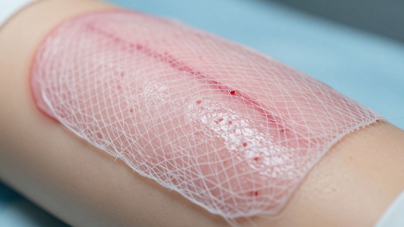
ECM as a biological scaffold rather than a passive covering
A passive covering mainly protects the wound from the outside. ECM participates more actively by supporting cell attachment, infiltration, communication, and remodeling. Its effect depends on the relationship between the material and the host wound, including the wound’s depth, moisture, inflammation, perfusion, and mechanical forces.
How collagen architecture supports epithelial cell movement
Collagen provides tensile structure and a framework that cells can grip as they move. Fiber arrangement, density, pore size, and degradation all affect whether cells can enter and cross the scaffold. The ECM cell migration literature describes this movement as a response to both physical architecture and biochemical guidance, not to collagen quantity alone.
The role of fibronectin, vitronectin, and other adhesion proteins
Fibronectin, vitronectin, periostin, and related proteins contain binding sites recognized by cell-surface receptors. These interactions help epithelial cells attach, spread, and generate traction. They also connect the structural matrix to intracellular signaling pathways that influence migration and proliferation.
How hydration and porosity influence cell activity
A hydrated matrix can support molecular transport and help prevent the wound surface from drying. Porosity affects fluid movement, cell entry, and the distribution of nutrients and signaling molecules. Too much fluid can cause maceration or dilute useful signals, while too little moisture can reduce cellular activity and make the surface less permissive to migration.
How ECM regulates epithelial cell behavior
ECM does not direct every cell in exactly the same way. Keratinocytes, fibroblasts, endothelial cells, and macrophages interpret matrix composition and architecture through different receptors and signaling pathways. The result is a local microenvironment in which cell behavior changes as the wound moves from inflammation toward repair.
Signals that attract epithelial cells into the wound
Chemokines, growth factors, and matrix-bound signaling molecules can create gradients that encourage cells to move toward damaged tissue. Epidermal growth factor and fibroblast growth factor are among the signals associated with epithelial activity and tissue repair, although their effects depend on dose, timing, receptor availability, and the condition of the wound. Matrix degradation can also expose new binding sites or release fragments that alter cell behavior.
Supporting cell attachment and spreading
A cell that cannot attach securely cannot migrate efficiently. Adhesion proteins and collagen domains allow epithelial cells to establish focal contacts, flatten, and organize their cytoskeleton. Stable cell attachment therefore serves as a practical bridge between a scaffold’s material properties and its biological effect.
Encouraging epithelial cell proliferation
Once cells have attached and received appropriate cues, proliferation increases the population available to cover the defect. Growth factors, matrix stiffness, nutrient availability, and signals from neighboring fibroblasts all influence this response. Excessive inflammation or an overly rigid, degraded, or poorly hydrated matrix can interrupt the transition from attachment to productive growth.
Coordinating migration across the wound bed
Migration works best when cells can follow a continuous, sufficiently stable path. A matrix with suitable fiber alignment and pore structure may help cells move in an organized direction rather than spreading randomly. That coordination matters because closure is not just about moving more cells; it is about moving them across the wound while maintaining a functioning, progressively maturing epithelial layer.
How ECM supports the other processes required for re-epithelialization
Epithelial closure depends on events beneath and around the surface. New blood vessels supply oxygen and nutrients, fibroblasts build provisional tissue, macrophages help regulate inflammation, and remodeling gradually changes the matrix into stronger tissue. If any of these processes remains severely impaired, the epithelial layer may be thin, unstable, or unable to advance.
Angiogenesis and oxygen delivery to regenerating tissue
Endothelial cells need a permissive matrix and appropriate signals to form new microvessels. Vascular ingrowth supports oxygen delivery, nutrient exchange, and removal of metabolic waste from the regenerating wound. ECM components such as glycosaminoglycans can also bind and organize growth factors involved in angiogenesis.
Fibroblast activity and formation of provisional tissue
Fibroblasts migrate into the wound, produce matrix, and contribute to granulation tissue. Their activity creates a more suitable base for epithelial cells while also influencing contraction and later scar formation. A scaffold should therefore be considered in relation to fibroblast behavior, not only its direct contact with keratinocytes.
Macrophage responses and the inflammatory environment
Macrophages help clear debris, coordinate inflammation, and support the transition toward repair. A prolonged inflammatory state can damage newly deposited matrix and maintain conditions that discourage epithelial migration. Research on cholecyst-derived matrices has investigated macrophage phenotypes and reparative host responses, but these findings should be interpreted as evidence of interaction with the material rather than proof that inflammation is eliminated.
Collagen remodeling and maturation beneath the new epithelium
New collagen is initially arranged in a provisional and relatively disorganized pattern. Remodeling changes fiber organization, turnover, and mechanical strength over time. The surface may close before this deeper maturation is complete, so ongoing protection and management remain important even after epithelial advancement becomes visible.
Why ECM composition and processing affect re-epithelialization
Two materials can both be described as collagen-based while behaving very differently in a wound. Tissue of origin, fiber architecture, associated proteins, hydration, processing, and degradation all influence how host cells respond. This is why evaluating the whole matrix is more informative than focusing on a single ingredient.
Why collagen alone may not reproduce a native healing environment
Collagen supplies structure and can support fibroblast activity, but native ECM contains many additional components that regulate adhesion, hydration, signaling, and remodeling. Removing those components may produce a simpler material with fewer biological cues. The distinction is explored further in this discussion of collagen beyond structure, which explains why collagen alone may not reproduce the full repair environment.
How elastin, glycosaminoglycans, proteoglycans, and growth factors contribute
Elastin supports flexibility, while glycosaminoglycans help retain water and organize signaling molecules. Proteoglycans such as decorin and lumican can influence collagen organization and cell behavior. Fibronectin, vitronectin, and growth factors add further cues for attachment, migration, angiogenesis, and proliferation.
A compact way to view these relationships is to separate the matrix into overlapping functions:
-
Collagens provide structural support and a surface for cell interaction.
-
Elastin and glycosaminoglycans contribute flexibility, hydration, and transport.
-
Proteoglycans help organize matrix structure and signaling.
-
Adhesion proteins support attachment, spreading, and migration.
-
Growth factors and matrix-bound molecules influence angiogenesis and proliferation.
This functional view helps you assess whether a scaffold offers more than bulk collagen. It also explains why preserving the relationships among components may matter as much as preserving the components themselves.
Effects of decellularization on ECM architecture and bioactive molecules
Decellularization aims to remove cellular material while retaining useful matrix structure and biomolecules. Harsh enzyme or detergent exposure may affect fiber organization or reduce biologically active components, although the outcome depends on the tissue and the validated process. The goal is not simply to remove cells, but to balance cellular clearance with preservation of the matrix features that host cells can use.
Balancing scaffold stability with controlled degradation
A scaffold must remain present long enough to support early repair, yet it should not persist indefinitely or prevent replacement by host tissue. Crosslinking and other stabilization approaches can alter strength and degradation rate, but they may also affect biological accessibility. The appropriate balance depends on wound conditions, intended use, and how the material interacts with the host over time.
What research suggests about cholecyst-derived ECM
Porcine cholecyst-derived ECM has been investigated as a naturally derived, collagen-rich scaffold with a three-dimensional fibrous architecture. The research includes structural characterization, protein profiling, cell interaction, immune response, degradation, and wound models. CholeDerm is discussed by Alicorn Medical in the context of a porcine-derived ECM approach, while the underlying research should still be read according to its specific model and formulation.
The mesh-like structure of porcine cholecyst-derived ECM
Published characterization describes a thin, flexible, porous, mesh-like collagen network. Such architecture may allow the scaffold to conform to a wound surface while providing spaces for fluid movement and cellular entry. Whether that structure performs well clinically still depends on wound preparation, application, exudate control, and the patient’s healing conditions.
Bioactive proteins associated with cellular attachment and migration
Protein-profiling work identified ECM-associated components including fibronectin, nidogen, decorin, and lumican, alongside collagen and other matrix constituents. These molecules are biologically relevant because they can influence adhesion, organization, and signaling. Their presence supports investigating cholecyst-derived ECM as a complex biological scaffold rather than treating it as processed collagen alone.
Findings from full-thickness wound and burn models
In rabbit full-thickness wound research, cholecyst-derived scaffold use was associated with reported granulation and earlier epithelialization in the evaluated model. A separate rabbit burn model reported reduced inflammatory findings and improved healing parameters, while other studies examined diabetic rat and dog wound models. These results suggest potential across several experimental settings, but each finding remains tied to its animal model, protocol, and formulation.
Interpreting preclinical evidence without overstating clinical outcomes
Animal studies can clarify material behavior, tissue response, vascularization, and remodeling before broader clinical evaluation. They cannot guarantee the same rate of closure, comfort, safety, or functional recovery for every person. You should therefore distinguish a study-reported preclinical result from a general product claim and consider human factors, wound cause, perfusion, infection, pressure, and comorbidities.
Applying ECM in a wound-healing strategy
ECM is one part of a wound-healing plan, not a replacement for diagnosis and wound-bed preparation. Before application, you need to assess tissue viability, perfusion, infection, pressure, exudate, depth, and the patient’s underlying conditions. The clinical guide to ECM wound application similarly emphasizes preparation, hydration, securing the matrix, moisture management, and correction of factors that may keep a wound from progressing.
Preparing the wound bed before ECM application
Remove nonviable tissue when clinically appropriate, address infection or bioburden, control bleeding, and confirm that the wound has a viable base. Adequate perfusion and pressure relief also matter because a biologic scaffold cannot compensate for untreated ischemia or repeated mechanical injury. Preparation should be individualized and performed according to clinical protocol.
A practical preparation sequence may include:
-
Assess wound depth, tissue viability, perfusion, infection, and exudate.
-
Debride or cleanse the wound as clinically indicated.
-
Control bleeding and address pressure, edema, or other mechanical problems.
-
Select a compatible ECM format and secondary dressing.
-
Establish a reassessment plan before application.
This sequence keeps the material within a broader treatment strategy rather than treating application as an isolated event. It also gives you clear points at which to reconsider the plan if the wound does not show meaningful progress.
Matching scaffold selection to wound depth and exudate
A thin, porous matrix may suit some superficial or full-thickness wound surfaces, but selection should reflect the geometry and exudate burden of the defect. Heavy drainage can interfere with contact and may require a secondary dressing or another method of fluid management. The material’s stability, handling, hydration requirements, and intended role should be checked against current product information and clinical judgment.
Combining ECM with dressings or negative pressure wound therapy
ECM can provide biological signals while a secondary dressing manages moisture and protects the wound. In selected high-exudate or complex wounds, negative pressure wound therapy may support fluid removal, wound contraction, and granulation alongside the scaffold. The knowledge base describes CholeDerm® with NPWT as a complementary approach in which the matrix serves as a biological interface and NPWT provides mechanical support; fenestration may be considered for fluid egress under an established protocol.
The PrisTINE™ process is described in Alicorn Medical materials as an enzyme-free, detergent-free approach intended to preserve native ECM architecture and biomolecules. That processing claim belongs to the documented process itself and should not be extended into assumptions about every wound or every clinical outcome.
Monitoring epithelial advancement and changing the treatment plan when needed
Monitor wound-edge advancement, tissue quality, exudate, odor, pain, maceration, bleeding, and signs of infection. Reassessment should also consider whether the underlying cause has been controlled, including pressure, vascular insufficiency, diabetes, or repeated trauma. If the wound is not improving, the dressing strategy and overall treatment plan should be reviewed with an appropriate clinician rather than continued automatically.
Conclusion
Understanding How ECM Supports Re-epithelialization means looking beyond surface coverage to the coordinated biology of attachment, migration, proliferation, vascularization, inflammation, and remodeling. A well-characterized ECM may provide structural and biochemical support, but its value depends on composition, processing, wound-bed preparation, patient factors, and careful clinical monitoring. Cholecyst-derived ECM research offers a useful preclinical example of how native architecture and matrix-associated biomolecules may influence repair while still requiring appropriately cautious interpretation.
Frequently Asked Questions
What is re-epithelialization?
Re-epithelialization is the process by which epithelial cells migrate, attach, proliferate, and spread across a wound to restore a continuous surface layer.
Why does ECM matter during re-epithelialization?
ECM provides physical attachment points and biochemical signals that help regulate epithelial migration, spreading, proliferation, and communication with other healing cells.
Is collagen enough to support epithelial repair?
Collagen is important, but native ECM also includes elastin, glycosaminoglycans, proteoglycans, adhesion proteins, and signaling molecules that contribute to a broader repair environment.
How can chronic wounds delay re-epithelialization?
Persistent inflammation, infection, poor perfusion, excess exudate, pressure, diabetes, and abnormal matrix degradation can interrupt the signals and conditions needed for epithelial advancement.
What role does angiogenesis play in epithelial closure?
New blood vessels deliver oxygen and nutrients, remove waste, and help sustain the cells and matrix involved in rebuilding the wound surface.
Can ECM replace wound-bed preparation?
No. Debridement when appropriate, infection management, perfusion assessment, pressure relief, exudate control, and treatment of underlying disease remain central to wound care.
What should you consider when interpreting ECM research?
Consider the tissue source, processing method, scaffold structure, wound model, formulation, follow-up period, and whether the evidence is preclinical or derived from human clinical use.