
Key Takeaways
ECM and Cell Migration During Wound Repair are closely connected: cells need both a path to follow and signals that tell them where to go. The quality of that local environment can shape whether repair progresses or stalls.
-
Cell migration brings immune cells, fibroblasts, endothelial cells, and keratinocytes into coordinated repair.
-
ECM structure provides attachment points, physical guidance, and biochemical signals.
-
Chronic inflammation, high protease activity, diabetes, ischemia, and infection can disrupt movement.
-
Biological scaffolds may support organized migration when their architecture and bioactivity are preserved.
-
Scaffold selection should follow wound-bed preparation, exudate control, vascular assessment, and clinical monitoring.
Why cell migration is essential for wound repair
Wound healing is not simply a matter of filling an empty space. You need successive waves of cells to enter the wound, communicate with one another, deposit matrix, form vessels, and restore the epithelial barrier. Migration links these events across the inflammatory, proliferative, and remodeling phases. A useful overview of this relationship appears in the discussion of ECM and wound healing, where matrix structure and cell behavior are considered together.
The role of migration in closing a wound
After hemostasis, cells move across and into the provisional wound matrix. This movement narrows the defect, supports new tissue formation, and brings repair cells into contact with signals from the wound bed. Without directional migration, proliferation alone cannot organize closure.
How acute and chronic wounds differ
An acute wound generally moves through inflammation and repair in a coordinated sequence. In a chronic wound, persistent inflammation, impaired angiogenesis, elevated matrix metalloproteinase activity, ischemia, or infection may keep the tissue in an unfavorable state. Cells may be present but move slowly, respond poorly, or encounter a matrix that is continually degraded.
Which cells migrate into the wound bed
Immune cells arrive early to clear debris and coordinate inflammation. Fibroblasts then enter to produce and remodel matrix, endothelial cells migrate during vascular growth, and keratinocytes move across the surface during re-epithelialization. Each population reads the wound microenvironment differently, so the same matrix can produce different cellular responses.
How migration supports granulation and re-epithelialization
Fibroblast migration helps build granulation tissue, while endothelial-cell movement supports capillary formation. Keratinocytes migrate from the wound edges to restore coverage and barrier function. Together, these processes turn a provisional matrix into living, vascularized tissue rather than a simple patch.
How the ECM guides migrating cells
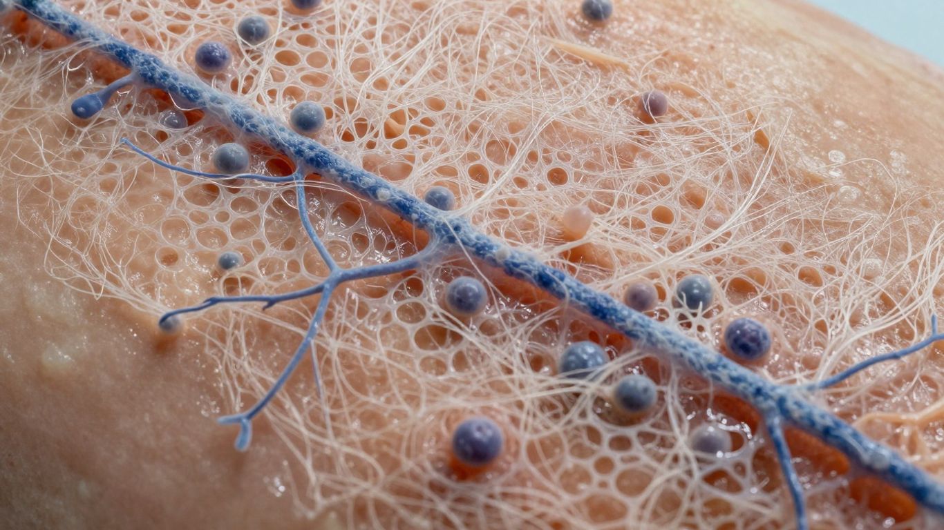
The extracellular matrix is both a physical environment and a source of information. Its fibers, pores, adhesion proteins, growth-factor binding sites, and mechanical properties influence how cells attach and move. This is why collagen alone does not describe the full biology; the wider ECM composition includes several components that coordinate migration and repair.
ECM as a physical scaffold
Cells need a surface that can support traction while they extend and retract their membranes. Collagen fibers and other matrix structures create routes through the wound bed, while pore size affects whether cells can enter, spread, or become constrained. A scaffold that is too dense may obstruct infiltration; one that is too loose may provide inadequate support.
Adhesion sites created by fibronectin and other proteins
Fibronectin, vitronectin, periostin, and related proteins create binding sites for cells. These sites let a cell grip the matrix, generate traction, and release that grip in a controlled sequence. The result is movement that is guided rather than random.
Integrins and cell–matrix communication
Integrins connect the ECM outside the cell to the cytoskeleton and signaling machinery inside it. When integrins bind matrix proteins, they can influence adhesion, spreading, migration, proliferation, and gene expression. The review on integrins in wound healing explains why these receptors are central to both granulation and re-epithelialization.
How matrix stiffness, porosity, and alignment influence movement
A cell responds to more than chemical composition. Stiffness changes traction, aligned fibers can create directional tracks, and interconnected pores affect infiltration. These features must be considered together because a matrix with useful proteins may still perform poorly if its geometry prevents cells from reaching the wound bed.
Chemotactic and haptotactic signals within the ECM
Chemotaxis describes movement toward soluble signals, while haptotaxis describes movement along gradients of matrix-bound cues. Growth factors can be retained by proteoglycans and glycosaminoglycans, creating local signals rather than a uniform bath. This combination helps recruit cells to the places where their work is needed.
ECM-driven migration across the stages of healing
Migration changes as the wound changes. Early signals recruit immune cells, the provisional matrix supports fibroblasts and endothelial cells, and later matrix remodeling enables epithelial closure and tissue maturation. You can therefore understand repair more clearly by following both the cells and the matrix through time.
Early inflammatory signals and immune-cell recruitment
Platelets and damaged tissue release signals that attract immune cells to the wound. These cells remove debris, regulate inflammation, and help prepare the matrix for repair. The goal is not to eliminate inflammation immediately, but to resolve it sufficiently for proliferative cells to take over.
Fibroblast migration during granulation tissue formation
Fibroblasts move into the wound and begin producing collagen, proteoglycans, and other matrix components. Their activity gives granulation tissue its structure and helps create a substrate for later epithelial movement. If the provisional matrix is poorly organized, fibroblast activity may become excessive or ineffective.
Endothelial-cell migration and new blood-vessel growth
Endothelial cells migrate toward angiogenic signals and form new vascular structures. This process supplies oxygen and nutrients to the growing tissue while also improving waste removal. Poor perfusion limits both the signals and the cellular energy required for migration.
Keratinocyte migration during re-epithelialization
Keratinocytes loosen their attachments at the wound edge, spread across the provisional matrix, and gradually restore the surface barrier. They need a moist, protected environment and a matrix that permits movement without excessive adherence. Delayed epithelial migration leaves deeper tissue exposed and increases the chance of complications.
Matrix remodeling and the transition to mature tissue
As repair progresses, enzymes remove some provisional matrix while fibroblasts deposit and reorganize new material. This controlled turnover changes the routes and signals available to cells. Remodeling must be balanced: too little leaves immature tissue, while too much can undermine the repair that cells have built.
When ECM structure and signaling fail
A wound can fail even when repair cells are present. Their movement depends on adequate perfusion, manageable inflammation, usable matrix signals, and a physical environment that is neither excessively wet nor desiccated. The chronic-wound ECM discussion describes how proteases and inflammatory cells can degrade the very proteins and growth factors needed for repair.
How chronic inflammation disrupts cell movement
Persistent inflammation changes the concentration of cytokines, proteases, and cellular debris in the wound. Instead of receiving a short, coordinated signal, migrating cells encounter repeated inflammatory stimulation. This can delay the shift toward granulation and epithelialization.
The effects of excessive matrix degradation and MMP activity
Matrix metalloproteinases are necessary for remodeling, but excessive activity can break down collagen, adhesion proteins, growth factors, and receptors. The wound then loses both its physical tracks and its biochemical instructions. Protecting a functional matrix requires addressing the cause of excess protease activity rather than simply adding more scaffold material.
Why diabetes and poor vascularization slow migration
Hyperglycemia, neuropathy, inflammation, and ischemia can interfere with cell function in diabetic wounds. Reduced blood flow limits oxygen delivery, while impaired angiogenesis slows the arrival of new vessels. These factors help explain why a diabetic wound may need metabolic, vascular, pressure, and infection management alongside local wound care.
Fibrotic ECM, tissue stiffness, and disorganized repair
A fibrotic matrix is often dense and mechanically abnormal. Excess stiffness can alter integrin signaling and encourage cells to deposit still more disorganized matrix. Repair may close the surface while leaving tissue with reduced flexibility and inferior architecture.
How infection and excessive exudate affect the wound microenvironment
Infection adds inflammatory burden and can damage newly formed tissue. Excess exudate may dilute signals, impair adhesion, macerate surrounding skin, and increase dressing challenges. Exudate control is therefore part of protecting the environment through which cells must migrate.
How biological ECM scaffolds support organized migration
A biological scaffold is most useful when it offers more than bulk collagen. Native matrix contains structural proteins, adhesion domains, glycosaminoglycans, proteoglycans, and signaling molecules that cells can interpret. The aim is to provide a temporary, compatible environment while the patient’s tissue repopulates and remodels it.
Preserving native architecture during scaffold preparation
Processing must remove cellular material while limiting unnecessary disruption of the matrix. Tissue source matters because a less cellular organ may require less aggressive processing than a highly cellular or intestinal source. When architecture is retained, fibers, pores, and molecular relationships remain closer to the original biological environment.
The contribution of collagen, elastin, GAGs, and proteoglycans
Collagen provides much of the structural framework, while elastin contributes flexibility. Glycosaminoglycans help regulate hydration and bind signaling molecules, and proteoglycans influence organization and growth-factor presentation. A broader explanation of GAGs in regenerative medicine helps clarify why these molecules matter for migration rather than serving as passive fillers.
Growth factors and bioactive molecules that influence migration
Growth factors such as VEGF and FGF can support angiogenesis and granulation, while adhesion proteins help cells attach and move. Their effect depends on concentration, presentation, and the surrounding matrix. Preserving several classes of biomolecules may therefore offer a more physiologic signal environment than adding one isolated factor.
Balancing scaffold integration with controlled degradation
A scaffold must remain long enough to support infiltration, but it must also permit replacement by new tissue. Stabilization or crosslinking can alter degradation, although excessive modification may affect bioactivity and cellular access. The right balance depends on tissue location, wound depth, mechanical demands, and the intended repair timeline.
Comparing natural ECM with collagen-only and synthetic materials
Different materials solve different design problems. The following comparison is useful when you are considering what a scaffold should contribute to a wound bed:
|
Material approach |
Main structural contribution |
Biological signaling |
Key consideration |
|---|---|---|---|
|
Natural ECM |
Multiple native matrix components and architecture |
Broad, tissue-derived cues |
Processing must preserve useful bioactivity |
|
Collagen-focused material |
Familiar structural protein and cell-supportive surface |
More limited molecular complexity |
May not reproduce the full native environment |
|
Synthetic scaffold |
Tunable geometry, strength, and degradation |
Often requires added bioactive cues |
Integration depends on surface and design |
The comparison is not a ranking for every wound. It shows why material selection should match the biological problem, rather than treating all scaffolds as interchangeable.
Cholecyst-derived ECM and cell migration research
Porcine cholecyst-derived ECM has been investigated as a naturally derived scaffold with collagen-rich, three-dimensional, mesh-like architecture. The research has examined composition, cellular interaction, tissue response, degradation, and wound repair in animal models. Commercial descriptions should remain distinct from research findings, so the evidence below is presented with its model and limitations intact.
The mesh-like architecture of porcine cholecyst-derived ECM
The reported cholecyst-derived scaffold has a thin, porous, tightly organized collagen network. Such a structure can provide a surface for cell attachment while allowing infiltration and fluid handling. Architecture is biologically relevant because it determines whether cells can enter, spread, and establish new tissue.
Evidence for fibroblast, endothelial-cell, and epithelial-cell interactions
The supplied studies report fibroblast attachment, endothelial-cell migration, and epithelial interaction with cholecyst-derived ECM. These observations are consistent with a scaffold that supports several coordinated stages of repair rather than one isolated cell type. They remain evidence from laboratory and preclinical work, not a guarantee of the same result in every patient.
How non-detergent and non-enzymatic processing may preserve bioactivity
A non-detergent and non-enzymatic recovery approach was investigated to reduce disruption of native matrix components. It should not be described as chemical-free, because stabilization and crosslinking conditions may still be used in scaffold preparation. The relevant question is whether processing clears cells while retaining architecture and useful biomolecules.
Findings from rabbit, diabetic-rat, and dog wound models
In rabbit full-thickness and burn-wound studies, the research reported granulation, epithelialization, and reduced inflammatory features in the evaluated models. A diabetic-rat study of a gelatin-modified cholecyst-derived scaffold reported enhanced angiogenic responses and faster healing in that model. A dog study of naturally occurring full-thickness lacerated wounds reported faster healing parameters for the cholecyst-derived scaffold group than the bovine dermal comparison.
These findings are best read as model-specific evidence. They support continued investigation of matrix architecture, angiogenesis, and host-material interaction, but they do not establish universal clinical outcomes.
Distinguishing preclinical evidence from clinical application claims
Animal studies can reveal how a scaffold interacts with tissue, how it degrades, and whether it supports organized repair. They cannot by themselves establish human efficacy, suitability for every wound, or a specific clinical protocol. For clinicians, the appropriate next step is to match current approved product information with wound assessment and the patient’s broader treatment plan.
Applying ECM principles to wound-care decisions
ECM science becomes clinically useful when it is connected to wound assessment. You first need to understand the wound’s cause, depth, perfusion, infection status, exudate level, pressure or mechanical forces, and patient comorbidities. A practical wound-bed preparation guide can help place scaffold use within debridement, hydration, fixation, offloading, compression, and monitoring rather than treating it as a stand-alone intervention.
Preparing the wound bed before introducing an ECM scaffold
Remove nonviable tissue when clinically appropriate, cleanse the wound, control infection, and assess perfusion before placing a biological scaffold. A clean, viable bed gives cells a better chance to attach and migrate. You should also address pressure, edema, glycemic control, and other causes that would continue damaging the repair environment.
Matching scaffold structure to wound depth and exudate level
A thin, porous scaffold may be useful where tissue infiltration and conformability matter, while a deeper or irregular wound may require additional strategies for space, coverage, and fluid management. Exudate should be controlled without drying the wound or flooding the matrix. Selection should follow the product’s approved instructions and the clinician’s assessment.
Combining ECM scaffolds with negative pressure wound therapy
Negative pressure wound therapy can help manage exudate, reduce edema, support granulation, and promote contraction in selected wounds. When used with a biological scaffold, the dressing interface and pressure settings must follow an appropriate clinical protocol. The advanced wound-care overview places NPWT among several adjuncts whose value depends on the wound’s cause and condition.
Monitoring granulation, epithelialization, and tissue integration
You can monitor whether the wound is becoming cleaner, whether granulation is progressing, whether epithelial edges are advancing, and whether exudate is becoming more manageable. Also watch for pain, odor, increasing drainage, bleeding, maceration, or other signs that the plan needs review. Serial photographs, measurements, and clinical documentation help distinguish steady progress from a temporary visual change.
Recognizing when vascular, infectious, or metabolic barriers require additional treatment
A scaffold cannot correct untreated ischemia, uncontrolled diabetes, significant infection, or ongoing pressure. A wound that fails to progress should prompt reassessment of perfusion, offloading, debridement, antimicrobial strategy, nutrition, and systemic disease. When the problem is complex, multidisciplinary wound care is often more valuable than simply changing the covering.
Conclusion
ECM and Cell Migration During Wound Repair depend on a coordinated relationship between cells, matrix architecture, biochemical signals, and the wound environment. By considering how cells attach, move, remodel, and respond to inflammation, you can evaluate biological scaffolds more thoughtfully and place them within complete wound care rather than viewing them as isolated dressings.
Frequently Asked Questions
What is the ECM in wound healing?
The extracellular matrix is a network of proteins, glycosaminoglycans, proteoglycans, and associated molecules surrounding cells. It provides structural support while also influencing adhesion, migration, signaling, inflammation, and remodeling.
Why is cell migration important for wound closure?
Migration brings immune cells, fibroblasts, endothelial cells, and keratinocytes to the areas where they are needed. These cells coordinate inflammation, granulation, vascular growth, and restoration of the epithelial barrier.
How does ECM stiffness affect migration?
Stiffness changes the traction cells can generate and can alter integrin signaling. Excessive or abnormal stiffness may redirect movement, increase fibrosis, or prevent cells from spreading in an organized way.
What is the difference between chemotaxis and haptotaxis?
Chemotaxis is movement along a gradient of soluble chemical signals. Haptotaxis is movement along a gradient of signals that are attached to the matrix or another surface.
Why do chronic wounds often heal slowly?
Chronic wounds may combine prolonged inflammation, high protease activity, impaired angiogenesis, ischemia, infection, neuropathy, pressure, and metabolic dysfunction. These conditions can damage matrix signals and make cell migration less effective.
Can an ECM scaffold replace wound-bed preparation?
No. Debridement when appropriate, cleansing, infection management, perfusion assessment, pressure relief, and control of systemic factors remain essential. A scaffold works within the wound environment and cannot remove every barrier to healing.
What should clinicians monitor after scaffold placement?
Monitor wound dimensions, granulation, epithelial advancement, exudate, odor, pain, tissue integration, and signs of infection or ischemia. Reassessment should guide ongoing treatment and confirm whether the wound is progressing as expected.